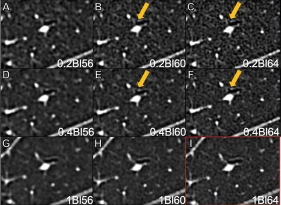
61-year-old male, post lung transplant, who underwent photon-counting detector CT of chest in ultra-high-resolution mode. Reconstructions were performed at 0.2-mm slice thickness with BI56 kernel (A), BI60 kernel (B), and BI64 kernel (C); at 0.4-mm slice thickness with BI56 kernel (D), BI60 kernel (E), and BI64 kernel (F); and at 1.0-mm slice thickness with BI56 kernel (G), BI60 kernel (H), and BI64 kernel (I). Clinical-reference reconstruction (BI64 kernel at 1.0-mm slice thickness) is shown with red outline in lower right (I). Small bronchus with wall discontinuity, corresponding to origin of bronchial division, is well visualized on reconstructions at 0.2-mm and 0.4-mm slice thickness with Bl60 and Bl64 kernels (arrows), but not on clinical-reference reconstruction. All images displayed with window of -400 HU and level of 1400 HU.
December 15, 2022 — An accepted manuscript published in ARRS’ American Journal of Roentgenology (AJR) guides optimization of clinical protocols when implementing ultra-high-resolution photon-counting detector (UHR PCD) CT of the lungs, providing insights on the association of reconstruction kernel and slice thickness with image quality.
Evaluating the impact of kernel and slice thickness on image quality of UHR PCD CT of the lungs using a 1024x1024 matrix, “the sharpest evaluated kernel, BI64, was the optimal kernel, consistent with the current clinical-reference technique,” wrote corresponding author Helmut Prosch from the department of biomedical imaging and image-guided therapy at the Medical University of Vienna in Austria.
In this AJR accepted manuscript, 29 patients (17 women, 12 men; median age, 56 years) underwent noncontrast chest CT using a first-generation PCD scanner (NAEOTOM Alpha, Siemens Healthineers, Forchheim, Germany) from February 15 to March 15, 2022. All acquisitions used UHR mode. Nine image sets were reconstructed for all combinations of three sharp kernels (BI56, BI60, BI64) and three slice thicknesses (0.2, 0.4, 1.0 mm). Three radiologists independently reviewed reconstructions for measures of visualization of pulmonary anatomic structures and pathologies using clinical-reference: BI641.0-mm.
Ultimately, when performing PCD CT of the lungs in UHR mode, reconstruction using BI64 kernel and 0.4-mm slice thickness was the only assessed reconstruction to yield improved bronchial division identification and bronchial wall and pulmonary fissure sharpness, without loss in pulmonary vessel sharpness or conspicuity of nodules or other pathologies.
In comparison, a 0.2-mm slice thickness—the thinnest reconstruction possible—was “associated with decreased visualization of various anatomic and pathologic findings,” the authors of this AJR accepted manuscript added.
For more information: www.arrs.org

 June 18, 2026


