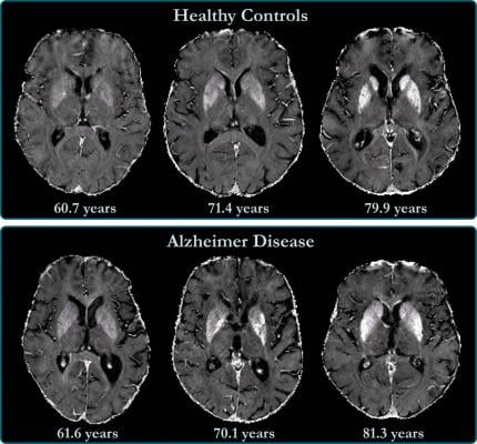
R2* maps of healthy control participants and participants with Alzheimer disease. R2* maps are windowed between 10 and 50 sec21. Differences in iron concentration in basal ganglia are too small to allow visual separation between patients with Alzheimer disease and control participants, and iron levels strongly depend on anatomic structure and subject age. Image courtesy of Radiological Society of North America
July 1, 2020 — Researchers using magnetic resonance imaging (MRI) have found that iron accumulation in the outer layer of the brain is associated with cognitive deterioration in people with Alzheimer's disease, according to a study published in the journal Radiology.
Alzheimer's disease is a progressive type of dementia that impairs and eventually destroys memory and other brain functions. There is no cure, although some treatments are thought to slow the progression.
Previous research has linked abnormally high levels of iron in the brain with Alzheimer's disease. Iron deposition correlates with amyloid beta, a protein that clumps together in the brains of people with Alzheimer's disease to form plaques that collect between neurons and disrupt cell function. Associations have also been found between iron and neurofibrillary tangles, abnormal accumulations of a protein called tau that collect inside neurons. These tangles block the neuron's transport system, which harms the communication between neurons.
It is known that deep gray matter structures of patients with Alzheimer's disease contain higher brain iron concentrations. Less is known about the neocortex, the deeply grooved outer layer of the brain that is involved with language, conscious thought and other important functions. The neocortex is challenging to assess by MRI, as the anatomy of the area makes MRI prone to distortions, signal decays and artifacts.
"The best solution to minimize these artifacts would be using ultra-high-resolution scans," said study coauthor Reinhold Schmidt, M.D., professor of neurology and chairman of the Department of Neurology at the Medical University of Graz in Graz, Austria. "However, in the clinical setting, scan time is a limiting factor, and a compromise has to be found."
For the new study, Schmidt and colleagues developed an approach using a 3T MRI scanner that allowed the best tradeoff between resolution and scan time, along with postprocessing to correct the influence of the distortions.
They used the MRI system to investigate baseline levels of brain iron in 100 individuals with Alzheimer's disease and 100 healthy controls. Of the 100 participants with Alzheimer's disease, 56 had subsequent neuropsychological testing and brain MRI at a mean follow-up of 17 months.
The technique enabled the researchers to create a map of brain iron, determining iron levels in parts of the brain like the temporal lobes, or the areas of the brain lying underneath the temples, and the occipital lobes in the back of the head.
"We found indications of higher iron deposition in the deep gray matter and total neocortex, and regionally in temporal and occipital lobes, in Alzheimer's disease patients compared with age-matched healthy individuals," Schmidt said.
The brain iron accumulation was associated with cognitive deterioration independently of brain volume loss. Changes in iron levels over time in the temporal lobes correlated with cognitive decline in individuals with Alzheimer's disease.
"These results are all in keeping with the view that high concentrations of iron significantly promote amyloid beta deposition and neurotoxicity in Alzheimer's disease," Schmidt said.
The results point to a potential role in Alzheimer's disease treatment for drugs that reduce the iron burden in the brain. These drugs, known as chelators, can remove excess iron from the body.
"Our study provides support for the hypothesis of impaired iron homeostasis in Alzheimer's disease and indicates that the use of iron chelators in clinical trials might be a promising treatment target," Schmidt said. "MRI-based iron mapping could be used as a biomarker for Alzheimer's disease prediction and as a tool to monitor treatment response in therapeutic studies."
For more information: www.rsna.org
Related Alzheimer's Imaging Content:
New Imaging Tool Helps Researchers See Extent of Alzheimer's Early Damage
Focused Ultrasound Opening Brain to Previously Impossible Treatments
Ultrasound’s Role in Alzheimer’s Treatment Research
Earlier Alzheimer's Diagnosis May Be Possible with New Imaging Compound

 July 20, 2026


