RELATED CONTENT

September 4, 2020 — According to ARRS' American Journal of Roentgenology (AJR), nearly half (47.7%) of the research ...
 September 04, 2020
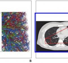
August 26, 2020 — An open-access article in ARRS' American Journal of Roentgenology (AJR) established a foundation for ...
August 26, 2020 
August 20, 2020 — According to ARRS' American Journal of Roentgenology (AJR), electronic consultation not only offered ...
August 20, 2020 
August 14, 2020 — According to ARRS' American Journal of Roentgenology (AJR), clinicians do not read a considerable ...
August 14, 2020 
February 12, 2020 — Mobile devices proved both reliable and accurate for the clinical decision to administer IV ...
February 12, 2020 
February 7, 2020 — An ahead-of-print article in the April issue of the American Journal of Roentgenology (AJR) reviewing ...
February 07, 2020 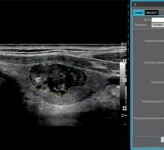
January 30, 2020 — According to an article published ahead-of-print in the April issue of the American Journal of ...
January 30, 2020 Although ultrasound remains the primary imaging modality used in prenatal imaging, fetal magnetic resonance imaging (MRI) is playing an increasing role in further evaluation of fetuses suspected of congenital anomalies. As 3-T MRI scanners become more common due to their improved image signal-to-noise ratio and anatomical detail, the benefits of 3-T MRI must be weighed against potential risks to the fetus that may result from the higher field strength.
April 22, 2015