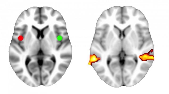
This image shows connectivity between the anterior insular cortex brain region (red and green spheres) and the middle temporal gyrus (orange-red gradient). Greater connectivity between these regions predicted better response to talk therapy for people with depression. Image courtesy of Andrew Crowther/Gabriel Dicther, Ph.D., UNC School of Medicine
February 6, 2015 — University of North Carolina (UNC) School of Medicine researchers have shown that brain scans can predict which patients with clinical depression are most likely to benefit from a specific kind of talk therapy.
The study, which was published in the journal Neuropsychopharmacology, is the first to use a technique known as resting-state functional brain connectivity magnetic resonance imaging (rs-fcMRI) to identify differences in brain wiring that predict therapeutic responses to talk therapy.
The research shows that brain scans could ultimately be used as a diagnostic tool to determine the best course of treatment for the millions of Americans that suffer from depression.
"In the future, we will be able to use non-invasive brain imaging technology to match patients with the treatment option that has the best chance of lifting their depression," said senior author Gabriel S. Dichter, Ph.D., associate professor of psychiatry and psychology. "In my mind, that's as important as developing new treatments. We already have a lot of excellent treatments but no way to know which one is best for a particular patient."
Dichter added that if doctors can identify the best treatment immediately, then doctors and patients could avoid months of trial and error, thus dramatically reducing the often-debilitating effects of depression for patients and their families.
Major depressive disorder, also known as clinical depression, is now the second leading cause of disability worldwide. Approximately 1 in 6 people will experience at least one bout of depression, and many will suffer multiple bouts over the course of their lives. Although antidepressant medications, different kinds of talk therapies and brain stimulation can be effective, 40 percent of people are not helped by the first treatment they try. As a result, Dichter said, it can take patients multiple attempts with different treatments before they experience any relief.
The researchers recruited 23 patients with major depressive disorder who were not yet being treated. The patients underwent rs-fcMRI, which visualizes the coordinated activity of various brain regions within known functional networks of neurons while the brain is not engaged in any particular tasks. By using this technique, the researchers could identify brain regions that light up or activate in unison. This, in turn, could help them uncover networks of activity that might be linked to certain behaviors or responses to therapy.
After the patients were scanned, they met with counselors for an average of 12 weekly talk therapy sessions using a method known as behavioral activation talk therapy. Whereas other forms of talk therapy might involve analyzing childhood experiences or altering thought processes, behavioral activation talk therapy focuses on the immediate behaviors associated with depression, such as difficulty getting to work on time or not spending time with loved ones. During the talk therapy sessions, patients set goals to address these behaviors.
Andrew Crowther, a graduate student in UNC's neurobiology curriculum and first author of the Neuropsychopharmacology paper, then analyzed the data to spot relationships between brain connectivity and responses to treatment. He found two connectivity patterns that stood out among patients who benefited most from talk therapy.
First, these patients had greater connectivity between the anterior insular cortex — a prune-sized region involved in assigning importance to events — and the middle temporal gyrus — a flattened section of brain tissue that plays a role in the subjective experience of emotion.
Second, patients had stronger connections between the intraparietal sulcus — a snake-like structure involved in maintaining focus — and the orbital frontal cortex — a crescent-shaped brain region behind the eyes involved in assigning positive or negative values to events.
"There's a complex interplay between the regions of the brain that are involved in cognitive control and those regions involved in understanding how something is going to feel," said Dichter, who is also a member of UNC's Carolina Institute for Developmental Disabilities.
"We've known for a long time that atypical connections between those regions are involved in depression, but now we know that they can also be involved in how a person responds to talk therapy."
Dichter and his colleagues plan to extend their imaging studies to explore responsiveness to other forms of talk therapy, anti-depressant medications, and brain stimulation.
For more information: www.med.unc.edu

 July 10, 2026


