News | January 12, 2015

Quantitative DTI scans can help solve concussion mysteries by showing a more detailed and complete image of the brain

MRI systems, Clinical Trial/Study, Diffusion Tensor Imaging

Image courtesy of Doctors Imaging


January 12, 2015 — Doctors Imaging offers a quantitative diffusion tensor imaging (DTI), a new MRI technology that can detect brain damage not appearing on routine MRIs or CT scans. Doctors Imaging is the only imaging facility in Louisiana, and one of only a few in the United States, offering quantitative DTI to better diagnose and ultimately treat concussions suffered in accidents, on the job or while playing sports.

“The need to diagnose concussions quickly and accurately has become a major issue in the medical community, especially with the prevalence of head injuries among athletes,” said Edward Soll, M.D., Doctors Imaging Concussion Program director.  “With the new quantitative DTI scans at Doctors Imaging, we can see evidence of brain injury that we weren’t able to see before and now can conclusively diagnose brain injury and better treat patients and their symptoms.”

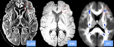
DTI can measure and evaluate small and subtle brain injuries by comparing a patient’s scan to a control group of normal subject DTI scans. This quantitative process helps to diagnose the specifically injured areas of the brain that can help physicians confirm the presence of nerve damage. A patient undergoing a quantitative DTI scan has a similar experience to a patient having a regular brain MRI scan, but the DTI results can reveal much more. As seen in the image below, the blue spots in the DTI brain scan on the right are nerve damage not seen in the two other standard scans of the same brain.

According to a study through the National Institutes of Health, the quantitative DTI scans now offered at Doctors Imaging can help solve those mysteries by showing a more detailed and complete image of the brain. Doctors Imaging has already successfully diagnosed patients whose conventional scans were negative.

For more information: www.doctorsimaging.com


Related Content

News | Magnetic Resonance Imaging (MRI)

Jan. 27, 2026 — Hyperfine has announced results from the largest data set to date evaluating stroke detection with its ...

Time January 28, 2026
arrow
News | Radiology Education

Jan. 22, 2026—The American Roentgen Ray Society (ARRS) will host a live virtual symposium, "Medical Imaging for ...

Time January 28, 2026
arrow
News | Radiology Imaging

Jan.26, 2026 — SimonMed Imaging has unveiled an updated brand and the launch of SimonMed Longevity, a new division ...

Time January 27, 2026
arrow
News | PET Imaging

Jan. 26, 2026 — Nuclidium, a clinical-stage radiopharmaceutical company developing a proprietary copper-based ...

Time January 27, 2026
arrow
News | Magnetic Resonance Imaging (MRI)

Jan. 20, 2026 — Hyperfine, the developer of the first FDA-cleared AI-powered portable MRI system for the brain — the ...

Time January 20, 2026
arrow
News | Stroke

Dec. 12, 2025 — Hyperfine, Inc. has announced that it has received FDA clearance for a new multi-direction diffusion ...

Time December 15, 2025
arrow
News | Artificial Intelligence

Dec. 1, 2025 — Researchers at the University of California, Berkeley and University of California, San Francisco have ...

Time December 10, 2025
arrow
Feature | Uzay Emir and Stephen Sawiak

Healthcare has reached a critical juncture. The World Economic Forum estimates that global medical costs will see double ...

Time December 04, 2025
arrow
News | Advanced Visualization

Nov. 20, 2025 — Avatar Medical and Barco have launched Eonis Vision, marking a new evolution in how medical imaging is ...

Time November 20, 2025
arrow
News | Neuro Imaging

Nov. 19, 2025 — Royal Philips has announced an extended partnership with Cortechs.ai. Together, the companies will ...

Time November 19, 2025
arrow
Subscribe Now