Researchers from University Hospitals Case Medical Center have published findings that PET/MRI is promising for several types of cancer.
September 3, 2013 — Researchers from University Hospitals Case Medical Center have published findings that a new form of imaging — magnetic resonance imaging/ positron emission tomography (PET/MRI) — is promising for several types of cancer. In an article titled “PET/MRI: Applications in Clinical Imaging,” published in the September issue of Current Radiology Reports, the authors outline their initial clinical experience in diagnosing and staging cancer patients with this novel technology.
Working in collaboration with researchers from Philips Healthcare, the team found that PET/MRI provided added value in the diagnosis, staging and treatment planning of colorectal cancers, cervical, uterine, ovarian and pancreatic cancers — as well as in the diagnostic management of pediatric and young adult patients. The researchers examined 145 cancer patients with a double-scanning protocol of PET/CT followed by a PET/MRI performed on the Philips Ingenuity TF PET/MRI system.
“Our preliminary experience with this new diagnostic imaging technology proves that it is promising for oncologic applications,” said study lead author Karin Herrmann, M.D., radiologist at UH Case Medical Center and visiting associate professor of radiology at Case Western Reserve University School of Medicine. “We found the PET/MRI enhanced our ability to detect malignant areas and more accurately and confidently diagnose several types of cancers, potentially providing physicians with the ability to improve treatment planning and better monitoring of the disease.”
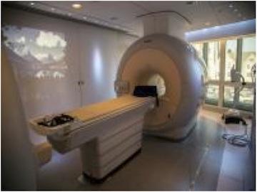
The PET/MRI is a new hybrid imaging modality which brings together the complementary capabilities of MRI scanning to better visualize both functional and anatomical information and to superimpose this information in a combined digital image. The UH Case Medical Center Seidman Cancer Center is the first cancer hospital in the world to house this technology in a purely clinical setting.
In a new class of diagnostic imaging modalities, the PET/MRI combines the highest anatomic detail as well as bio-chemical and functional information provided by MRI with the metabolic, molecular and physiologic information from PET. The technology fuses the images to more precisely pinpoint cancer locations and improve the accuracy of disease staging.
In the study, the authors’ detailed account summarizes many aspects of the clinical utility of PET/MRI in more accurate detection of cancer and cancer metastasis, use in decision-making, comparisons of detection accuracy across cancers and distinction of benign from malignant lesions in clinical settings.
The authors also outline the considerations of reduction in overall radiation imaging exposure with PET/MRI versus other imaging technologies. Given the similar performance of PET/MR compared to PET/ CT in some disease entities, there is potential to decrease radiation exposure in replacing the CT component in PET/CT with MRI. This may especially be an issue in pediatric and young adult patients with need for repetitive follow up imaging.
“This hybrid scanner has the potential to improve patient care by increasing understanding of the causes, effects, and development of disease processes to better diagnose cancer and various other diseases,” said study author Norbert Avril, M.D., nuclear radiologist at UH Case Medical Center and professor at Case Western Reserve University School of Medicine. “We are very excited to be among the first to be able to help establish guidelines of how best to use this technology to guide physicians on the value of the PET/MRI in diagnosing and staging various forms of cancer. Our initial experience has shown that it may be a very important cancer-fighting tool.”
This research was supported by Philips Healthcare and is part of the Philips Healthcare Global Advanced Imaging Innovation Center, established in 2009 at UH Case Medical Center and Case Western Reserve to bring the latest Philips Healthcare imaging equipment to Cleveland for development, validation of clinical efficacy and product release. The Imaging Center was funded through an initial $5 million from Ohio Third Frontier Commission to Case Western Reserve University School of Medicine and a $33.4 million matching commitment from Philips.
The Department of Radiology at UH Case Medical Center and Case Western Reserve University School of Medicine, and the Department of Biomedical Engineering at Case Western Reserve School of Engineering, have some of the nation’s leaders in medical imaging who partner with Philips. The company’s global computed tomography and nuclear medicine headquarters is also in Cleveland.
“The collaboration between our various organizations has created a pipeline to move innovative technologies — such as the PET/MRI — more quickly into patient care,” said Pablo Ros, M.D., chairman of the Department of Radiology at UH Case Medical Center and Case Western Reserve School of Medicine. “Through this center, we have become an international hub for imaging technology and there has been great synergy between our institutions to make great strides through imaging research like this to help patients with cancer as well as heart disease and neurologic conditions.”
For more information: www.uhhospitals.org

 July 10, 2026


