The more you know about a disease, the better you can characterize it and treat it, and the path toward personalized medicine is clearly mapped out in PET imaging.
Positron emission tomography (PET) has already proven effective in helping clinicians diagnose, stage, treat and monitor many types of tumors and lesions in the body, and yet its utility continues to expand across more cancers and diseases.
As the development of novel biomarkers and drug discovery using PET systems opens new avenues to detection and treatment, experts predict that the use of PET will increase by an estimated 120 percent over the next 10 years. With no end to the number of applications in sight, the question is how far will this technology bring physicians down the road of personalized medicine?
FDG-PET makes its mark
Several clinical oncology programs today enable early assessment of tumor status using (18-fluoro-deoxy-glucose) FDG-PET. Currently, researchers at the National Cancer Institute (NCI) are investigating the use of FDG-PET as a potential biomarker for clinical trials conducted in non-Hodgkin’s lymphoma and non-small cell lung cancer. The FDG-PET lung and lymphoma clinical trials, which emerged from the Oncology Biomarker Qualification Initiative (OBQI), and the hope is that it will streamline drug approval.
“We anticipate that the findings of the FDG-PET Lymphoma Project will result in a 20-week reduction in the time it will take for the FDA to approve new lymphoma drugs and eventually other cancer drugs as well,” indicated Deborah Banker, Ph.D., vice president, Research Communications, The Leukemia & Lymphoma Society.
PET’s role as a biomarker opens the door to new opportunities in cancer care. “Although we are early in the development of imaging tools as biomarkers, this research holds the potential, over time, to be used not only in the diagnosis of cancer, but in monitoring and predicting response to therapy,” noted John Niederhuber, M.D., director, National Cancer Institute.
In prostate cancer, for example, researchers presented a study at the American Society of Clinical Oncology (ASCO 2007)1, in which they assessed the value of FDG-PET imaging as a biomarker of systemic therapeutic response for patients with prostate cancer bone metastases. They found that early findings suggest changes in FDG and 11C-acetate PET uptake describe prostate cancer bone metastasis response to systemic therapy.
PET can also be an effective tool for early prediction of response to chemotherapy. A recent study revealed how PET effectively monitored the cell cycle response of both tumor and bone marrow to cytotoxic chemotherapy.2 Following treatment with gemcitabine, tests revealed an early flare in uptake of 3'-deoxy-3'flurothymidine (FLT), a PET tracer, in many tumors. In contrast, uptake in bone marrow was reduced. Researchers found that in patients with metastatic colorectal cancer (mCRC) sequential FDG-PET predicted progression-free survival (PFS) and was more accurate than clinical response criteria.
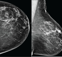
New strides in breast cancer appear to be gaining ground after a new study released this year indicated that PET might be more effective than mammography and ultrasound in breast treatment response. Researchers measured FDG uptake in tumors from patients with locally advanced breast cancer before and after preoperative chemotherapy. Women who had the highest accumulation at the beginning and who then had the highest percentage drop in accumulation after four cycles of chemotherapy were more likely to have a complete response to their treatment. However, measurements taken using mammography or ultrasound were not able to predict a pathological response accurately.
“FDG-PET appears to be an important addition to conventional imaging of women with breast cancer and may contribute significantly to a more individualized management of their disease,” said Vinod Ganju, M.D., Monash Oncology Research Institute (MORI) and Monash Breast Cancer Research Consortium, Monash Medical Centre, Melbourne, Australia.
PET/CT – one-stop shop
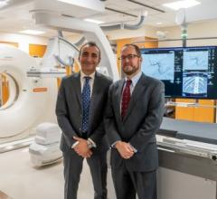
One of the biggest strides in PET technology has been the change from the stand-alone PET system to the hybrid PET/CT system. This has enabled the computed tomography (CT) to add the missing anatomical information to functional data captured by PET.
Referring to the PET/CT system, Joe Busch, M.D., the Diagnostic PET/CT Center of Chattanooga in Tennessee calls it “a one-stop shop for the cancer patient. Traditionally, we did a diagnostic CT and then that patient came back for a PET scan on another day or week. Now, if you have a an abnormality on a chest X-ray, that patient can go straight for the diagnostic CT and the PET scan simultaneously. It saves patient time and expedites the treatment. To me, it’s better patient care.”
The one-stop-shop concept can also apply to expediting image-guided biopsies. “We do PET/CT-directed biopsies. We biopsy the hot spot, which is unique,” said Dr. Busch. “It expedites patient care because you have them on the table, you see the abnormality, you execute the biopsy, if it is necessary.” Dr. Busch believes that in the future, more sites will use diagnostic CT simultaneously with PET to save the patient two exams.
Paradigm shift in cardiac imaging
PET/CT is also gaining importance in nuclear cardiac imaging. By adding the CT component to PET, the system provides a faster way to produce cardiac images and obtain additional information about coronary calcium deposits due to atherosclerosis.
“By identifying the metabolic correlate of an anatomical lesion, hybrid PET/CT can differentiate benign from malignant lesions,” said Vasken Dilsizian, M.D., FACC, FAHA, professor of Medicine and Radiology, chief, Division of Nuclear Medicine at the University of Maryland Medical Center. “However, the combined assessment of coronary artery lesions (anatomic information) with downstream functional consequence (blood flow to the heart muscle) of such anatomic lesions has not received widespread clinical acceptance in cardiology at the present time. Given that stand-alone PET systems are being largely replaced with hybrid PET/CT systems, driven predominantly by oncology, it is likely that such combined assessment of anatomy and physiology will flourish further in cardiology as well.”
Current advances in technology with hybrid PET/CT will allow for a paradigm shift, Dilsizian believes, from detection and treatment of coronary artery disease to prediction and prevention of coronary artery disease by detecting coronary atherosclerosis early in its progression. According to Dr. Dilsizian, this would provide a strong rationale with objective evidence for monitoring the disease progression, which would be particularly relevant among those at high risk for coronary artery disease such as diabetes mellitus.
“Given the noninvasive nature of PET/CT, one could potentially apply this technique for screening rather than diagnosing coronary artery disease,” he indicated. “The earlier one can identify and halt the atherosclerosis process, the better the patient outcome. This could potentially decrease the incidence of sudden cardiac deaths as well.
As PET technology advances rapidly today, what lays ahead in the future? Clinicians like Ronald D. Petrocelli, M.D., chief medical officer, Siemens Molecular Imaging, hope it will lead to very personalized therapy. “We’re striving for individualized therapy,” said Dr. Petrocelli. “Then we will see development of drugs specific to that type of tumor.” If this vision of customized drug development one day becomes a reality, we will likely be seeing it through PET.
Feature | November 14, 2007 | Cristen C. Bolan
Oncology is only the beginning as new PET applications open the road to personalized care across multiple diseases.
© Copyright Wainscot Media. All Rights Reserved.
Subscribe Now


 February 09, 2026


