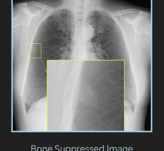
February 13, 2015 — XRware is launching an advanced new image processing software called XRware Virtual Grid. The included technologies improve image quality for exams that are taken without a grid.
The system automatically adapts its processing to replicate grid use to reduce degradation of image quality caused by scatter radiation, with no user interaction required.
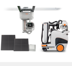
Physical add-on grids are commonly required for mobile imaging of large anatomy to help focus radiation to reduce scatter and blurring. XRware Virtual Grid processing is a benefit for mobile imaging in emergency rooms, operating rooms, nursing homes, bedsides, wheelchairs and other exams where the optimal grid or any grid may not be on-hand. The system also eliminates artifacts typical from misalignment of the tube to detector/grid angle.
The clinician will benefit as it will offer better visibility and contrast of the small and weak structure, better depiction of overlapping structure, simultaneous viewing of bone and soft tissue, reduced window width & window leveling and improved motion unsharpness reduction.
XRware Virtual Grid is available as a software development kit (SDK) available for integration into an existing product.
Part of the XRware software suite, the Virtual Grid offers the following seven modules and specifications:
- XRware Simulation for whole-system modelling for a more accurate system design, XR control setup and system calibration. Features include X-ray spectrum simulation, detector response simulation, interaction X-ray/matter, system uncertainty modeling, dose estimation
- XRware 2-D Image Processing to experience image processing for a more accurate analysis of images. Features include noise reduction, shading correction, sinogram-based processing, contrast enhancer, high dynamic range imaging, flux variation, projection registration, multi-energy processing.
- XRware Tomography Reconstruction to generate object model from its projections using artifact-reduced approaches using whatever trajectories and acquisition constraints (partial scan, low dose acquisitions). Features include analytical approaches, exact reconstruction algorithm, approximated reconstruction algorithms (FDK/FBP, BFP), acquisition strategies (circular, non-circular, helix, complex trajectories), extended view, partial scan, iterative approaches (deterministic and stochastic approaches), automatic Hounsfield unit calibration, automatic geometry parameters adjustment.
- XRware 3-D Image Processing to experience volume processing for a more accurate analysis. Features include noise reduction, contrast enhancement, sharpening, ring artifact reducer, beam hardening reduction, metal artifact reduction, scatter reduction
- XRware Analysis, a range of tools aiding quantification and diagnosis
Other features include
- Segmentation
- Measurement/metrology
- Mesh extraction
- CAD comparison
- Volume comparison
- Porosity analysis
- Data fusion
- XRware Acquisition features include acquisition from most popular digital radiography (DR) manufacturers
- Visualization and accurate data representation tools for a better visualization study
- Volume rendering
- Mesh rendering
- 2-D/3-D representation
- Hierarchical visualization
- LUT management
For more information: www.xrware.com

 February 10, 2026
