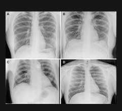

December 16, 2015 — Siemens Healthcare announced the U.S. Food and Drug Administration (FDA) has cleared the Multitom Rax robotic advanced X-ray. The Multitom Rax is a universal diagnostic imaging system that enables a wide range of examinations in multiple clinical areas – from emergency medicine and interventional to pain management and orthopedics, and from conventional 2-D radiography to fluoroscopy examinations and angiography applications – all in one room using one X-ray system. The world’s first Twin Robotic X-ray system, according to the company, the Multitom Rax features a unique design that enables, for the first time, the acquisition of 3-D natural weight-bearing images.
The open design of the system features a height-adjustable patient table and two independent, ceiling-mounted robotic arms for the X-ray tube head and the flat-panel detector for almost unlimited positioning freedom anywhere in the room. Both robotic arms can be moved into position automatically or manually with servo motor support to make fine adjustments. While one robotic arm moves the X-ray tube, the other arm carries the 17” x 17” flat panel detector, which can acquire static, dynamic and Real 3-D sequences. The operator is always in control of the system’s movement and able to position both robotic arms precisely and safely around the patient.
The Multitom Rax enables, for the first time, the acquisition of 3-D images under the patient’s natural weight-bearing condition – whether they are seated, lying down or standing. Images acquired in the natural standing position are essential because the knees, pelvis and spinal column appear differently when the patient’s body weight is applied compared to when the patient is lying down. Currently, 3-D imaging on the Multitom Rax is only possible with 3-D-capable post-processing software.
The Multitom Rax’s 3-D-ready image capabilities can offer better diagnostic and planning certainty than images that do not reflect a natural weight-bearing condition. Conventional 2-D X-rays do not always reveal fine hairline fractures in the bone. In cases of a suspected bone fracture, the patient historically has required a computed tomography (CT) 3-D image to confirm the diagnosis. With the Multitom Rax Twin Robotic X-ray system, a 3-D image can be acquired on the same system, so the patient does not have to wait for a future appointment or be transferred to a CT system.
Built-in features like Comprehensive CARE (Combined Applications to Reduce Exposure) and more precise positioning of the system aid in reducing patient radiation dose.
For more information: www.siemens.com

 October 07, 2026