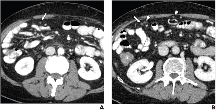
66-Year-Old Man With Locally Advanced Pancreatic Cancer Undergoing Chemotherapy: (A) Axial image from contrast-enhanced CT using positive oral contrast material, performed to monitor treatment response. Examination clinically interpreted as not showing metastatic disease. Unblinded retrospective image review shows tiny omental nodule (arrow) near bowel loops. Adequacy of bowel filling with contrast material rated very good. Both blinded retrospective readers detected nodule. (B) Axial image from contrast-enhanced CT performed two months later shows slight increase in size of nodule (arrow) and several new nodules (arrowheads), confirming lesion as missed malignant deposit. Image courtesy of American Roentgen Ray Society (ARRS), American Journal of Roentgenology (AJR)
April 27, 2022 — According to ARRS’ American Journal of Roentgenology (AJR), the selection of oral contrast agent and optimization of bowel preparation for oncologic computed tomography (CT) could help avoid potentially severe clinical consequences of missed malignant deposits.
“CT has suboptimal NPV for malignant deposits in intraabdominal nonsolid organs,” wrote corresponding author Benjamin M. Yeh of the University of California, San Francisco. “Compared to neutral material, positive oral contrast material improves detection, particularly with adequate bowel filling.”
Yeh and team’s retrospective study included 265 patients (133 men, 132 women; median age, 61 years) who underwent an abdominopelvic CT examination where the report did not suggest presence of malignant deposits and subsequent CT examination within 6 months where the report indicated at least one unequivocal malignant deposit. Examinations used positive (iohexol; n=100) or neutral (water; n=165) oral agents. While reviewing images to assess visibility of deposits, a board-certified abdominal radiologist also assessed adequacy of bowel filling with oral contrast material.
NPV of CT for detection of malignant deposits in intraabdominal nonsolid organs was 65.8% for examinations using positive oral contrast material with adequate bowel filling, 45.2% for positive oral contrast material with inadequate bowel filling (p=.07), and 35.2% for neutral oral contrast material regardless of adequacy of bowel filling (p=.002).
“Results may differ when studying other types of contrast material regimens,” the authors of this AJR article noted, including barium-based, hyperosmolar iodine, sugar-alcohol neutral, or experimental dark oral agents.
For more information: www.arrs.org

 June 15, 2026


