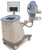
Naviscan PEM scanner
May 12, 2010 - Positron emission mammography (PEM) may reduce unnecessary breast biopsies, according to new data from an National Institutes of Health (NIH)-sponsored, multisite study of hundreds of women with newly diagnosed breast cancer.
The study found that PEM had a high positive predictive value (PPV) in identifying benign and cancerous lesions, and therefore may reduce the number of unnecessary biopsies.
This finding is a welcomed outcome for women and physicians looking for ways to reduce the patient trauma associated with biopsies and for payors looking to reduce the costs associated with unnecessary procedures.
The NIH-sponsored multisite study (NIH Grant 5R44CA103102) examined women with newly-diagnosed breast cancer. Patients were accrued from six leading clinical centers across the country: ARS Johns Hopkins Green Spring, Boca Raton Community Hospital, Scripps Clinic-Scripps Green Hospital, University of North Carolina, University of Southern California Norris Cancer Center, and Anne Arundel Medical Center.
The 388-woman study showed that PEM not only demonstrated a six percent improvement in specificity at comparably high sensitivity, but that PEM also had 31 fewer unnecessary biopsies and 26 percent higher PPV than breast magnetic resonance imaging (MRI). These results are also particularly significant for those women who cannot tolerate an MRI exam and require an alternate imaging tool.
“The results of this study mean that not only do physicians have an additional, powerful tool to help treat breast cancer but that PEM is a legitimate and better alternative for the 16 percent of women who cannot tolerate MRI due to claustrophobia, metallic implants, body habitus, or gadolinium reaction,” said Wendie Berg, M.D., Ph.D. and principal investigator for the trial.
PEM scanners are high-resolution breast positron emission tomography (PET) systems that can show the location as well as the metabolic phase of a lesion. This information helps determine whether a lesion is malignant and influences the course of treatment. Other imaging systems, such as mammography and ultrasound, show only the location, not the metabolic phase.
For more information: www.naviscan.com

 July 07, 2026


