Getty images
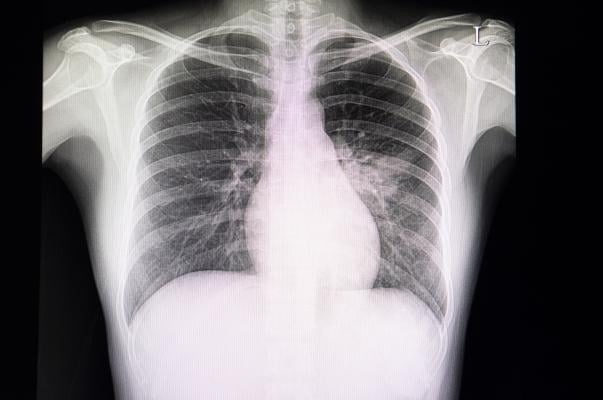
April 14, 2020 — Chest X-rays are often used to detect infections in the lungs, but the world’s largest study of its kind finds it’s not a reliable way to diagnose respiratory infections caused by COVID-19.
The research team led by Michael Weinstock, M.D., an adjunct professor of emergency medicine at The Ohio State University College of Medicine, reviewed more than 630 chest X-rays of confirmed and symptomatic COVID-19 patients of a large urgent care company in New York and New Jersey. The radiologists determined the chest x-rays were normal in 58.3% of cases, and normal or only mildly abnormal in 89% of patients.
“Providers ordering a chest x-ray in the outpatient setting should be aware that a patient with symptoms of COVID-19 may have a negative chest X-ray and should manage the patient based on their symptoms,” said Weinstock. “Doctor’s should not be reassured by a negative chest X-ray.”
Additionally, the research in The Journal of Urgent Care Medicine found the chest x-ray results of COVID-19 patients differ from those of bacterial pneumonia patients. The abnormal chest x-ray findings of COVID-19 patients were more likely to be scattered diffusely throughout both of the lungs than typical pneumonia.
“This study reinforces what we’ve learned from our colleagues outside of the United States that the majority of patients with COVID-19 have mild symptoms and minimal evidence of disease on chest X-ray,” said Matthew Exline, M.D., associate professor in the department of Pulmonary, Critical Care and Sleep Medicine at The Ohio State University College of Medicine and medical director of the medical intensive care unit at The Ohio State University Wexner Medical Center. “Hopefully this will help clinicians decide who would best benefit from hospitalization and any potential new treatments.”
Researchers are working on a second study to look at the outcomes of these patients and how it correlates to their chest X-ray results.

 December 10, 2025