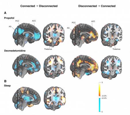
Differences in brain activity between connected and disconnected states of consciousness studied with positron emission tomography (PET) imaging. Activity of the thalamus, anterior (ACC) and posterior cingulate cortices (PCC), and bilateral angular gyri (AG) show the most consistent associa-tions with the state of consciousness (A = general anesthesia, B = sleep). The same brain struc-tures, which are deactivated when the state of consciousness changes to disconnected in general anesthesia or natural sleep (cool colors in the left columns), are reactivated when regaining a con-nected state upon emergence from anesthesia (warm colors in the right columns). Graphic courtesy of University of Turku
December 30, 2020 — What happens in the brain when our conscious awareness fades during general anesthesia and normal sleep? Finnish scientists studied this question with novel experimental designs and functional brain imaging. They succeeded in separating the specific changes related to consciousness from the more widespread overall effects, and discovered that the effects of anesthesia and sleep on brain activity were surprisingly similar. These novel findings point to a common central core brain network fundamental for human consciousness.
Explaining the biological basis of human consciousness is one of the greatest challenges of science. While the loss and return of consciousness, as regulated by drugs or physiological sleep, have been employed as model systems in the study of human consciousness, previous research results have been confounded by many experimental simplifications.
"One major challenge has been to design a set-up, where brain data in different states differ only in respect to consciousness. Our study overcomes many previous confounders, and for the first time, reveals the neural mechanisms underlying connected consciousness," said Harry Scheinin, Docent of Pharmacology, Anesthesiologist, and the Principal Investigator of the study from the University of Turku, Finland.
A new and innovative experimental set-up
Brain activity was measured with positron emission tomography (PET) imaging during different states of consciousness in two separate experiments in the same group of healthy subjects. Measurements were made during wakefulness, escalating and constant levels of two anesthetic agents, and during sleep-deprived wakefulness and Non-Rapid Eye Movement (NREM) sleep.
In the first experiment, the subjects were randomly allocated to receive either propofol or dexmedetomidine (two anesthetic agents with different molecular mechanisms of action) at stepwise increments until the subjects no longer responded. In the sleep study, they were allowed to fall asleep naturally. In both experiments, the subjects were roused to achieve rapid recovery to a responsive state, followed by immediate and detailed interviews of subjective experiences from the preceding unresponsive period. Unresponsive anesthetic states and verified NREM sleep stages, where a subsequent report of mental content included no signs of awareness of the surrounding world, indicated a disconnected state in the study participants. Importantly, the drug dosing in the first experiment was not changed before or during the shift in the behavioral state of the subjects.
"This unique experimental design was the key idea of our study and enabled us to distinguish the changes that were specific to the state of consciousness from the overall effects of anesthesia," explained Annalotta Scheinin, Anesthesiologist, Doctoral Candidate and the first author of the paper.
Researchers discovered a common central core brain network
When PET images of responsive and connected brains were compared with those of unresponsive and disconnected, the scientists found that activity of the thalamus, cingulate cortices and angular gyri were affected independently of the used anesthetic agent, drug concentration and direction of change in the state of consciousness (see figure). Strikingly analogous findings were obtained when physiological sleep was compared with sleep-deprived wakefulness. Brain activity changes were much more extensive when the disconnected states were compared with a fully awake state. State-specific findings were thus distinct and separable from the overall effects of drug-induced anesthesia and natural sleep, which included widespread suppression of brain activity across cortical areas.
These findings identify a central core brain network that is fundamental for human consciousness.
"General anesthesia seems to resemble normal sleep more than has traditionally been thought. This interpretation is, however, well in line with our recent electrophysiological findings in another anesthesia study," saidHarry Scheinin.
Subjective experiences are common during general anesthesia
Interestingly, unresponsiveness rarely denoted unconsciousness (i.e., total absence of subjective experiences), as most participants reported internally generated experiences, such as dreams, in the interviews. This is not an entirely new finding as dreams are commonly reported by patients after general anesthesia.
"However, because of the minimal delay between the awakenings and the interviews, the current results add significantly to our understanding of the nature of the anesthetic state. Against a common belief, full loss of consciousness is not needed for successful general anesthesia, as it is sufficient to just disconnect the patient's experiences from what is going on in the operating room," explains Annalotta Scheinin.
The new study sheds light on the fundamental nature of human consciousness and brings new information on brain functions in intermediate states between wakefulness and complete unconsciousness. These findings may also challenge our current understanding of the essence of general anesthesia.
For more information: www.

 July 23, 2026


