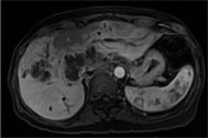
Figure 1: Arterial phase 3-D VIBE FS, PAT 2, 25 second breath-hold, 3 mm, 180 x 320 matrix, 25 x 34 cm
Frank Miller, M.D., medical director of magnetic resonance (MR) at Northwestern Memorial Hospital Outpatient Imaging Center in Chicago, Ill., shares a case in which he imaged complex liver masses on Siemens’ 1.5 Tesla Magnetom Aera and 30-channel body coil combination.
Case History
A 51-year-old female with a history of malignancy of unknown primary and elevated liver function tests.
Techniques: Axial/coronal sub-second T2 HASTE, axial in/out of phase breath-hold T1 FLASH, axial T2FS, axial diffusion, axial/coronal pre/post contrast breath-hold 2-D T1 FLASH; axial/coronal thick slab 1.5 second HASTE for thick slab MRCP and coronal free-breathing 3-D T2 SPACE for 3-D MRCP.
Case Summary
Evaluation of the liver demonstrates several ill-defined infiltrative masses which demonstrate progressive peripheral enhancement on delayed phase sequences with lack of central enhancement (see Figure 1).
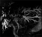
There is marked intra-hepatic ductal dilatation associated with these lesions which has progressed from the prior study. The dilation is best visualized by the 3-D SPACE sequence (see Figure 2, below) acquired in a free-breathing acquisition without belts or bellows. An automated phase navigator sequence synchronizes data acquisition to the patient’s breathing for high quality.
The gallbladder is surgically absent with susceptibility artifact noted in the gallbladder fossa.
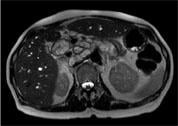
Massive conglomerate lymphadenopathy is noted involving the porta hepatis, mesenteric and retroperitoneal regions (see Figure 3, below).
Impression:
1. Interval progression of ill-defined infiltrative hepatic masses from metastatic disease.
2. Intrahepatic biliary ductal dilatation.
3. Massive conglomerate lymphadenopathy within the upper abdomen appears grossly stable.
About Siemens 70 cm open bore systems
As the innovator of 70 cm open bore design, Siemens Healthcare delivers MRI systems that allow you to take maximum advantage of the 70 cm bore. The standard, fully digital RF system offers 48 RF channels and 204 coil elements to create an imaging matrix that allows maximum use of coil elements at full FoV without ever running out of RF channels.
Siemens' unique Tim coils allow more flexible scanning positions. Not all patients, such as those with compromised breathing, kyphosis or Paget’s disease, can lie flat on their backs during an MRI exam. With Tim coils, you have the freedom and flexibility to use unique scanning positions (e.g., on a patient’s side) and still obtain the image quality you need for high clinical confidence. In addition, Tim allows full use of parameters (iPAT, Fat Sat, diffusion, etc.) in these positions for high-quality images that provide referring physicians with the information they need to manage their cases.
Aera: Take your guided tour today
Skyra: Take your guided tour today
For more information: www.medical.siemens.com

 July 02, 2026


