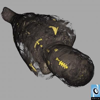
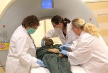
June 29, 2010 – Researchers were able to use 21st-century technology to learn more about a mummy from the 18th century last week using a noninvasive computerized tomography (CT) scan. The scan, which was completed at the Cedars-Sinai Medical Center’s S. Mark Taper Foundation Imaging Center, will help determine the mummy’s state of preservation and any disease or injury he may have had.
The adult male mummy, Michael Orlovits, and his two mummy family members are on loan from the Hungarian Natural History Museum, Budapest, as part of the “Mummies of the World” exhibition, the largest collection of mummies and related artifacts ever assembled, making its world debut July 1 at the California Science Center in Los Angeles.
“CT scans and other science tools represent the gold standard in studying mummies, helping us to learn much more about how people lived and died,” says Dr. Heather Gill-Frerking, director of science and education for the Mummies of the World exhibition and the scientific research curator for the German Mummy Project. “These techniques are also noninvasive and provide a complete three-dimensional archive record, which also allows us to preserve the mummies for future generations.”
Orlovotis and his family are part of a group of 18th-century mummies discovered in Vac, Hungary, in 1994. Reconstruction of parts of a Dominican church just north of Budapest uncovered two long-forgotten burial crypts dating back to 1674 and sealed in 1838.
Michael Orlovits, Veronica Orlovits (born 1770) and their son Johannes (born 1800) were among those preserved by the cool, dry air of the crypt and the oil from the pine shavings that lined some of the coffins. Extensive research, including DNA analysis, revealed that Veronica Orlovits suffered from severe tuberculosis. The scan conducted at Cedars-Sinai on Michael Orlovits will help reveal if he suffered from the same disease, in addition to any other diseases or injuries. Without invasive techniques, the scan also will reveal the exact condition of preservation of the mummy over the past 245 years.
“Mummies of the World” is making a three-year, seven-city tour around the country beginning in Los Angeles at the California Science Center on July 1.
For more information: www.mummiesoftheworld.com.

 June 18, 2026


