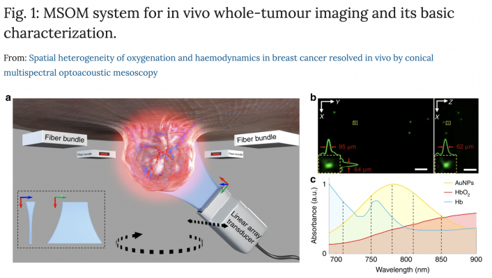
a Schematic of the system. The entire solid tumour is illuminated from four sides by a four-arm fibre bundle. A cylindrically focused linear array is designed to detect optoacoustic signals from the tumour. In vivo imaging is performed in conical scanning geometry by controlling the rotation and translation stages. The sensing part of the transducer array and the tumour are submerged in water to provide acoustic coupling. b Maximum intensity projections of the optoacoustic reconstruction of a phantom of polyethylene microspheres (diameter, 20 μm) dispersed in agar. The inset shows a zoomed-in view of the region boxed with a yellow dashed line. In addition, the yellow boxes are signal profiles along the x, y and z axes across the microsphere centre, as well as the corresponding full width at half-maximum values. c Normalized absorption spectra of Hb, HbO2 and gold nanoparticles (AuNPs). The spectrum for the AuNPs was obtained using a USB4000 spectrometer (Ocean Optics, Dunedin, FL, USA), while the spectra for Hb and HbO2 were taken from http://omlc.org/spectra/haemoglobin/index.html. The vertical dashed lines indicate the five wavelengths used to stimulate the three absorbers: 710, 750, 780, 810 and 850 nm. Optoacoustic signals were filtered into a low-frequency band (red) and high-frequency band (green), which were used to reconstruct separate images.
May 26, 2020 — Breast cancer is the most common cancer in women. But individual tumors can vary significantly, presenting different spatial patterns within their mass. Researchers at the Technical University of Munich (TUM) and Helmholtz Zentrum München have now succeeded in visualizing spatial changes within tumors by means of optoacoustics. This method may be helpful for the future development of new drugs.
Malignant tumors consume nutrients and oxygen faster than healthy cells. To do so, they recruit blood vessels in their environment. Depending on tumor type and genetic profile, there are differences on how tumors look internally. Typically, tumors present different patterns across their volume. The role of this spatial heterogeneity is not well understood or studied in living tumors. Typically used to understand biological functions in tumors, optical microscopy, for example, gives limited insights into the spatial heterogeneity of tumors as it only accesses volumes of less than a cubic millimeter.
High resolution with new imaging method
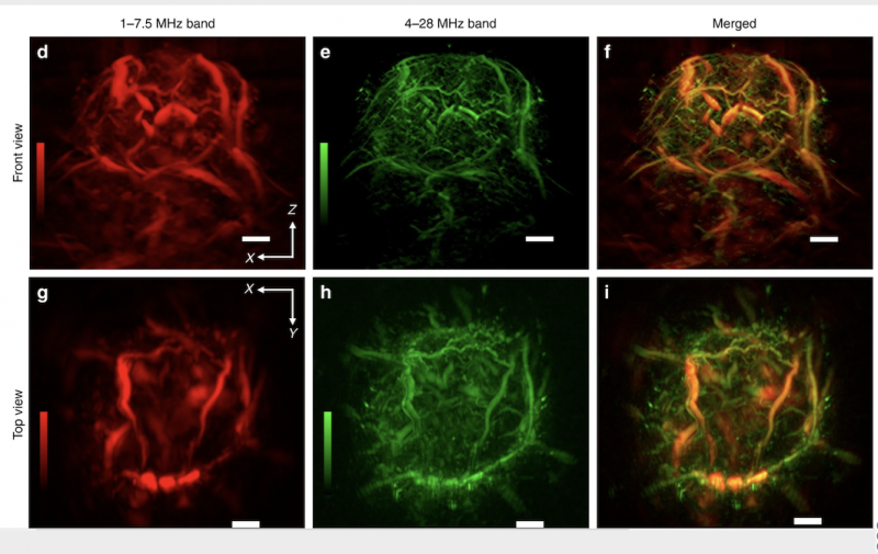
A new technique developed by Munich researchers, known as multi-spectral optoacoustic mesoscopy (MSOM), has now been shown capable of resolving optical contrast through tumor volumes that are at least 1,000 times larger than those possible with optical microscopy, enabling high-resolution visualization of tumor heterogeneity patterns. With this imaging method, the tumor is first excited from all sides with pulses of infrared laser light. “Tumor and tissue components that absorb this excitation light undergo a tiny, transient temperature increase, which leads to a small local volume expansion, followed by a contraction. This expansion and contraction process generates a weak ultrasound signal, which we collect with a detector,” said guest researcher Jiao Li, M.D.
The data collected is mathematically processed to form light absorption images which indicate different tumor patterns reflecting tumor oxygenation and vascularization. “For the first time, MSOM offers optical images that reach inside tumors to depths of ten millimeters and more with a resolution of less than 50 micrometers,” said Li.
Understanding functional variety in tumors
“MSOM imaging of solid tumors allowed us to see tumors in a new light,” said Prof. Vasilis Ntziachristos, holder of the Chair of Biological Imaging at TUM and Director of the Institute for Biological and Medical Imaging at Helmholtz Zentrum München. “MSOM allows us to understand how tumor functionality varies across the tumor, clearly moving the reach of optical observations well beyond the depth penetration limitations of optical microscopy.”
In the pictures taken from mamma carcinomas of mice, researchers can see patterns indicating the presence or absence of blood vessels, and thus study blood supply patterns. MSOM can also resolve hemoglobin levels, and indicate whether oxygen is bound to hemoglobin or not. Furthermore, MSOM images were used to determine permeability of the vessel walls relative to nanoparticles. Using the mouse model, the scientists were already able to track how tiny gold particles were transported.
3-D tumor pictures without surgical biopsy
In contrast to conventional histology, where tissue has to be removed, cut up and examined under the microscope by a pathologist, MSOM allows a three-dimensional analysis of entire living tumors without the need for surgical biopsies. This further supports longitudinal studies so that tumor growth or recession under different drugs can be studied with greater accuracy. All of which paves the way for an improved understanding of biological function and drug efficacy during drug development for humans.
For more information: www.tum.de/

 July 07, 2026


