March 19, 2019 — Late-breaking results confirm the HeartFlow FFRct (fractional flow reserve computed tomography) Analysis enables efficient identification of which patients, despite symptoms suggestive of coronary artery disease (CAD), have a low risk of adverse cardiovascular events and can safely avoid invasive testing out to one year. These results from the ADVANCE trial were presented as a late-breaking trial during the American College of Cardiology’s (ACC) 68th Annual Scientific Session, March 16-18 in New Orleans, and simultaneously published in the Journal of the American College of Cardiology (JACC): Cardiovascular Imaging.
In the ADVANCE study of more than 5,000 patients, clinicians used FFRct values from the HeartFlow Analysis to help determine patients’ risk of adverse cardiovascular events and decide upon management plans. The vast majority of the patients who had a negative HeartFlow Analysis received medical therapy and did not receive invasive testing or treatment. At one year, patients in this group had a significantly low rate (0.2 percent) of cardiovascular death or heart attack (myocardial infarction). In comparison, the rate of cardiovascular death or heart attack was four times higher in patients with a positive HeartFlow Analysis, many of whom required invasive management. These results demonstrate that patients identified as lower risk for adverse events may be safely treated with medications alone and avoid invasive management. Importantly, these patients had low rates of revascularization (stenting or bypass surgery) through 90 days and negligible need for revascularization thereafter.
“These findings provide reassurance regarding the safety of patient management utilizing an FFRct-guided decision pathway, particularly in lower-risk patients who did not undergo an invasive evaluation,” said Manesh Patel, M.D., FACC, chief, Division of Cardiology, Department of Medicine, Duke University School of Medicine, Duke University Health System. “By adding the HeartFlow FFRct to our available resources for diagnosing stable coronary disease, we are able to provide patients with better care as we efficiently evaluate risk in patients getting a coronary CTA, more precisely stratify patients and improve efficiency in the cath lab.”
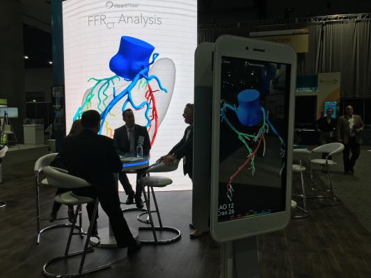
The HeartFlow Analysis is a non-invasive, cardiac test for stable symptomatic patients with CAD, the leading cause of death worldwide. Starting with a standard coronary computed tomography angiogram (CTA), the HeartFlow Analysis creates a digital, personalized 3-D model of the heart and provides FFRct values along the coronary arteries. This information helps physicians evaluate the impact a blockage may be having on blood flow and determine the best treatment for each patient. A positive FFRct value (≤0.80) indicates that a coronary blockage is impeding blood flow to the heart muscle to a degree which may warrant invasive management.
In the ADVANCE Registry, patients from the United States, Japan, Europe and Canada underwent a coronary CTA, and when additional information was needed, a HeartFlow Analysis was ordered. The added information contained in the HeartFlow Analysis led physicians to reconsider and change management plans for two-thirds of their patients. Some who were originally scheduled to receive a coronary stent or bypass operation were safely able to avoid the procedure and be treated with medications alone, while others who would have received medications were redirected to stenting or bypass surgery.
For more information: www.heartflow.com

 July 15, 2026


