

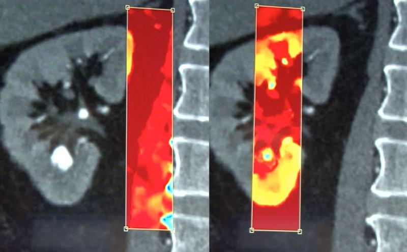
IQon allows use of a spectral window that can be moved around the image to show the chemical makeup of tissue in the frame. Here it shows the composition of a kidney stone with regular CT imaging and spectral.

November 26, 2014 — Philips Healthcare has received U.S. Food and Drug Administration (FDA) 510(k) clearance for its IQon Spectral computed tomography (CT) technology for spectral imaging. It adds a new dimension to CT imaging, delivering anatomical information and offering the ability to characterize structures based on their material content within a single scan. The enhanced image quality may help improve confidence in diagnoses and deliver operational efficiency.
Philips IQon Spectral CT was developed in close collaboration with clinicians and is designed to overcome some of their most immediate challenges, including workflow and image management issues. The technology can discriminate between X-ray photons of multiple high and low energies — similar to the principal behind a prism splitting white light into the rainbow of colors. Through the Spectral CT scan, clinicians can access the conventional grayscale anatomical images, along with the spectral information within the same scan. As a result, Philips IQon Spectral CT enables real-time, retrospective data analysis without disrupting a clinician's workflow and takes the guesswork out of multi-energy acquisitions, making it easy to use and allowing for routine spectral use.
The spectral CT is on all the time, so nothing needs to change in the clinical workflow. The spectral data can be accessed after a scan to help differentiate materials such as metal and bone to help reduce beam hardening. Spectral data can also help differentiate calcified arterial plaque or kidney stones based on the chemical composition of materials. It can also reconstruct a virtual non-contrast CT scan from contrast enhanced images by filtering out the iodine based on the spectral data.
"Spectral imaging has the potential to change the way clinicians practice radiology in the future," said Jacob Sosna, president of the Israeli Radiology Society and chairman of the department of radiology at Hadassah Hebrew University Medical Center, who collaborated with Philips on the development of the product. "A recent study1 with Philips IQon Spectral CT revealed enhanced diagnosis in up to 70 percent of cases. Using the prospective approach, we would only have access to spectral information in 20 or 25 percent, at maximum."
The IQon Spectral CT system is currently available for ordering in 36 countries. Philips will be showcasing the IQon Spectral CT at RSNA 2014.
"Delivering an additional level of spectral information without impacting clinician workflow is vital for confident diagnoses that can significantly add value to help improve patient care," said Gene Saragnese, CEO imaging systems, Philips Healthcare.
For more information: www.philips.com/healthcare
Reference:
1. Gabbai, M, et al. "The Clinical Impact of Retrospective Analysis in Spectral Detector Dual Energy Body CT." Radiological Society of North America 2013 Scientific Assembly and Annual Meeting, December 1 - December 6, 2013, Chicago IL. http://archive.rsna.org/2013/13018312.html Accessed June 24, 2014.


 June 18, 2026


