April 7, 2010 - A dual-modality tomographic (DMT) breast scanner, developed by researchers at the University of Virginia Health System (UVA), in a pilot study pinpointed to a much finer degree than mammography, ultrasound, MRI and even a needle biopsy, the location of breast masses and more accurately distinguished between cancerous and harmless lesions.

The pilot clinical study, led by Mark B. Williams, Ph.D., associate professor of radiology, biomedical engineering and physics at the University of Virginia, appears in the April 2010 issue of Radiology.

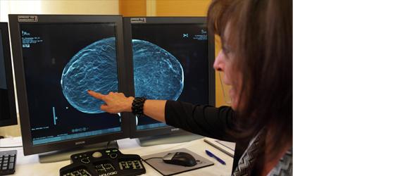
The DMT breast scanner works by coupling 3D digital X-ray breast tomosynthesis with 3D molecular breast imaging tomosynthesis, a technique that uses intravenously injected compounds (tracers) that are absorbed to a much greater degree by malignant lesions than benign ones. A special camera positioned in close proximity to the breast then performs functional imaging of the entire breast, while the digital X-ray tomosynthesis obtains co-registered structural images. The device runs the scans sequentially, obtaining both types of images with the breast in the same, immobilized position.

According to the researchers, the scanner particularly useful for women with radiographically dense breasts. Williams points out, this pilot clinical study of the 17 women tested could have far greater implications.

For more information: www.healthsystem.virginia.edu


Related Content

News | Breast Imaging | Washington University

Jan. 22, 2026 — In breast cancer, a biopsy is the only diagnostic procedure that can determine if a suspicious lump or ...

Time January 29, 2026
arrow
News | Radiation Oncology

Jan. 27, 2026 — Researchers at the Icahn School of Medicine at Mount Sinai, in collaboration with other leading ...

Time January 29, 2026
arrow
News | Radiology Education

Jan. 22, 2026—The American Roentgen Ray Society (ARRS) will host a live virtual symposium, "Medical Imaging for ...

Time January 28, 2026
arrow
News | Computed Tomography (CT)

Jan. 21, 2026 — Aidoc recently announced that the U.S. Food and Drug Administration (FDA) cleared the industry's first ...

Time January 23, 2026
arrow
News | Point-of-Care Ultrasound (POCUS)

Jan. 22, 2026 — Qure.ai has received a grant from the Gates Foundation to develop a large open-source multi-modal ...

Time January 23, 2026
arrow
News | Radiology Imaging

Jan. 21, 2026 — Cathpax, a spin-off of the Lemer Pax group that designs, develops and commercializes team-wide, full ...

Time January 22, 2026
arrow
News | PACS

Jan. 21, 2026 — Fujifilm Healthcare Americas Corp. and Voicebrook, Inc. have announced a strategic partnership to ...

Time January 22, 2026
arrow
News | Radiology Education

Jan. 20, 2026 — The American Society of Radiologic Technicians (ASRT) Foundation has named ASRT member Danielle McDonagh ...

Time January 20, 2026
arrow
News | Mammography

Jan. 16, 2026 — Vega Imaging Informatics has announced the successful curation of the world’s largest digital breast ...

Time January 19, 2026
arrow
News | Breast Imaging

Jan. 14, 2026 — VizMark has received U.S. Food and Drug Administration FDA 510k clearance for VM1, a non-metal visual ...

Time January 19, 2026
arrow
Subscribe Now