
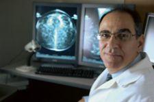
\"The most important aspect of the digital image is that it can be manipulated by the radiologist,\" said Dr. Semine.
The overriding philosophy guiding the entire approach to patient care for the Women’s Imaging Center of Newton-Wellesley Hospital (NWH) is the need to create a comprehensive medical environment where a woman can receive all of the services she needs in one location and in one day.
For Newton-Wellesley, located in the suburbs of Boston, this comprehensive approach allows patients to receive any necessary follow-up procedures quickly. “If a woman comes in for a mammogram, and we find she needs additional views, an ultrasound, or a biopsy, we’ll do it during the same visit,” said Alan Semine, M.D., chief of Breast Imaging. “The time frame in which a diagnosis is rendered makes a big difference to the patient.”
Making the Digital Transition
One of the reasons Dr. Semine came to the Imaging Center was because NWH understood the value of digital imaging and was committed to moving into the digital world. “All of radiology is becoming digital,” Dr. Semine said, “and it is important that mammography departments also benefit from the potential of the digital environment.”
Dr. Semine knew that if they were going to make the transition, and make it work for the patient, it was going to take time to integrate the new systems into the existing workflow. “We didn’t want to change the way we approached patient care,” he said. “Going digital before we had the proper processes in place was a serious concern.
“Hologic was very receptive when we suggested early on in our digital mammography journey that we would need some type of electronic communication between radiologist and the mammographer obtaining additional views. Because of this the Hologic technologist workstation was developed,” Dr. Semine said. NWH has opted to install technologist workstations in each exam room as well as one in the mammography work area. This allows the mammographer who is taking additional views to review the images in the exam room – similar to bringing analog images that have been circled into the exam room for review.
Leap of Faith
After months of research, including site visits and meetings with technology vendors and medical professionals, the decision was made to move ahead with the purchase of the Hologic Selenia digital mammography technology. “We were confident that we would go with Hologic,” Dr. Semine said, “because the image quality is really the best on the market.”
“We looked at all the vendors, and what they could bring to our patients here,” said Rich Guarino, Director of Radiology at NWH. “We bought the Selenia system based on image quality, and how that quality improves patient care.”
In fact, the Center was so impressed with the quality of the digital images from the Selenia that it committed to the purchase of the first unit on the contingency that the Hologic Selenia Technology received approval from the FDA.
“Our workflow has improved as a direct result of the Selenia technology,” said Deborah Lockhart, Operations Manager of Women’s Imaging. “We’re no longer printing images or burning CDs, thus increasing the efficiency within the department.”
According to Dr. Semine, the digital images from the Selenia system are far superior to analog film systems. “The most important aspect of the digital image is that it can be manipulated by the radiologist,” he said. “You’re able to interact with it much more effectively and bring out features that you never could with analog.”
The radiologists of the Women’s Imaging Center, in association with Hologic, recently completed a study in the use of complementary imaging software to enhance still further the detail in the images generated by the Selenia DM system. “Providing physicians with the technology to see more detail with better clarity is quite valuable,” Dr. Semine said. “Improving the display of visual information inherent in digital mammograms can ultimately help physicians detect breast cancers more effectively.”
The Women’s Imaging Center has found that the digital environment also allows for a seamless integration of their Hologic R2 Computer Aided Detection (CAD) software, improving the image interpretation workflow for their radiologists. “I worked with CAD since it first became available,” Dr. Semine said. “Once it’s been incorporated into the workflow, it proves to be valuable and makes even more of a difference in the digital environment.”
Working Through the Growing Pains
With the implementation of any new technology, a certain amount of growing pains are expected, and the first Selenia system installed in the Center was no exception. What made all the difference is that Hologic understood this and worked closely with the doctors, administrators and technologists at the Center to improve the new technology and integrate their suggestions into subsequent updates.
One improvement was the need for better communication between the radiologist and the imaging technologist at the acquisition station. “We worked closely with Hologic to validate the concept of a technologist workstation and demonstrate the improvement in workflow,” Dr. Semine said.
The Hologic technologist workstations allowed the Selenia technology to imitate, in a way, the analog environment. “We can now circle areas in the breast on the digital image that we would like the technologist to provide additional views of,” Dr. Semine said, “and push it right back to the technologist’s workstation. This has improved the efficiency of the digital environment.”
According to Breast Imaging Facilitator Cheryl Cain, the Selenia technology would not be as impressive if it did not improve the delivery of care to the patients. “We want to be hi-tech and on the cutting edge of technology; the Selenia has enabled us to do that,” Cain said. “But it’s all for patient care. Doing the best for the patient is the bottom line.”
A key factor affecting the decision to go with Hologic was its ability to accommodate different breast sizes easily. Selenia offered not only a large detector but also a shifting compression paddle that enables accurate positioning and imaging of small breasts “One of the reasons we decided to go with Selenia was the larger detector,” said Lockhart. “The size of the detector was important in our decision.”
The larger detector means that women with larger breasts can be imaged in one exposure, more easily. “We really like the ability to care for all patients regardless of size,” said Cain, “and without having to change image receptors. The compression paddle is all you need to switch.”
Fully Digital
After a four-year transition, Women’s Imaging Center is now fully digital, boasting a total of seven Selenia digital mammography systems, three dedicated radiologist diagnostic workstations and eight technologist workstations.
The new digital workflow is allowing the Women’s Imaging Center to improve the patient experience for a growing number of women. In fact, since beginning the transition to digital, the Center has seen an increase in annual patient volumes averaging 20 percent per year, for each year, since the first Selenia system was installed. “We want to be able to continue growing, but we don’t want our growth to have a negative effect on the quality of care that women have come to expect at the Center. What attracts patients here is our way of taking care of them,” said Dr. Semine.

 June 02, 2026


