October 16, 2009 - Researchers utilizing computed tomography (CT) scans have found that patients with severe cases of the H1N1 virus are at risk for developing severe complications, including pulmonary emboli (PE), according to a study published online Oct. 14, 2009, in the American Journal of Roentgenology.
In the study, Chest Radiographic and CT Findings in Novel Swine-Origin Influenza A (H1N1) Virus (S-OIV) Infection, which was published online at www.ajronline.org on October 14, and will appear in the December issue of the AJR, researchers set out to review the chest radiographic and CT findings in patients with presumed/laboratory-confirmed novel swine-origin influenza A (H1N1) virus (S-OIV) infection.
The study, performed at the University of Michigan Health Service, included 66 patients diagnosed with the H1N1 flu. Two study groups were formed. Group one consisted of 14 patients who were severely ill and required Intensive Care Unit (ICU) admission. Group two consisted of 52 patients who were not severely ill and did not require ICU admission.
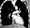
All 66 patients underwent chest X-rays for the detection of H1N1 abnormalities. Ten patients from the ICU group and five patients from the largely outpatient group, underwent CT scans. "Pulmonary Emboli were seen on CT in five of 14 ICU patients," said Prachi P. Agarwal, M.D., lead author of the study.
The researchers concluded that chest radiographs were normal in more than half of patients with S-OIV (H1N1) and progress to bilateral extensive air-space disease in severely ill patients, who are at a high risk for PE.
Reference: Agarwal, Prachi P. Radiographic and CT Findings in Novel Swine-Origin Influenza A (H1N1) Virus (S-OIV) Infection. AJR:193, December 2009 1. American Journal of Roentgenology.
For more information: www.ajronline.org

 June 18, 2026


