June 7, 2011 — Research presented at SNM’s 58th annual meeting is taking targeted molecular imaging to a new level by combining two commonly used imaging agents into one molecular imaging procedure. The combination of these agents creates a comprehensive examination of the extent of cancer spread within a variety of organ systems in the body.
“During a time when healthcare costs are under intense scrutiny, consolidated procedures such as this one that provide comprehensive imaging data are a benefit to everyone — to clinicians, healthcare administrators and especially patients who would need only one scan instead of two,” said Andrei Iagaru, M.D., lead author of the study and assistant professor of radiology and nuclear medicine at Stanford University Medical Center, Stanford, Calif. “What we stand to gain from combining these two agents is the diversification of tumor targeting. The beauty of it is that each agent has its own strength, and those are unified in the imaging. In combination they represent a powerful new tool for acquiring as much information as possible about the extent of a patient’s cancer.”
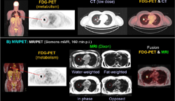
The objective of the study was to gauge the value of combining two molecular imaging agents for cancer imaging with a hybrid molecular imaging system. Researchers used a positron emission tomography (PET)/computed tomography (CT) system, which combines both nuclear medicine and X-ray technology to produce images that provide information about both physiological processes and structural anatomy.
All PET imaging agents are not alike. Medical isotopes are often labeled with an antibody, peptide or any number of other useful molecules that guide the medical isotope directly to its target. One of the most prevalently used imaging agents is 18F-fluorodeoxyglucose (18F FDG), a glucose analog that is metabolized by cells as fuel. This is useful because cancer cells are metabolically hyperactive compared to healthy cells. PET imaging with 18F FDG will show these areas of increased metabolism as “hot spots” on scans. But while it is an excellent agent, it can still miss some cancer lesions, such as those in the skeleton. For this study, researchers compare the results of 18F FDG and sodium fluoride-18 (18F NaF), another PET imaging agent that has been shown to provide superior functional imaging of skeletal tissue, both separately and together.
For this prospective study, investigators recruited 78 patients with proven cancers and scanned them separately using 18F NaF PET/CT and 18F FDG PET/CT imaging methods. Participants then received a third PET/CT scan using 18F NaF and 18F FDG simultaneously. Results of all three imaging studies were then analyzed for their usefulness in evaluating tumor malignancy. Researchers found that combined 18F NaF and 18F FDG PET/CT imaging provided good-quality imaging of both skeletal and extra-skeletal malignant lesions from breast, lung, colorectal and other cancers.
Combined 18F NaF and 18F FDG PET/CT is a comprehensive new imaging procedure for cancer staging without any additional risk of toxicity. In fact, this technique reduces overall radiation dose due to the consolidation of imaging. With further demonstration of its clinical value, this imaging technique could become widely available for evaluating a host of different cancers in order to improve treatment planning and patient prognosis.
For more information: www.snm.org

 July 15, 2026


