March 12, 2010 – A new advanced visualization technology in development allows clinicians to automatically apply supercomputing algorithms for the analysis of internal organs, vessels, tumors and other regions of interest in multiple dimensions, from 2D to 5D, including data set animations. Ziosoft Inc. displayed its work-in-progress PhyZiodynamic solutions for its Ziostation system at the annual American College of Cardiology (ACC) Scientific Sessions March 14–16 in Atlanta.
“PhyZiodynamic technology provides imaging solutions that are truly remarkable compared to traditional volumetric rendering,” said Sung Woo Park, M.D., Ph.D., professor of the department of medicine, Cardiac and Vascular Center, Samsung Medical Center, Sungkyunkwan University School of Medicine. “When I realized I could clearly see the biomechanics of the papillary muscles, it was apparent there was something very significant here. I believe this technology has the potential to offer a substantial impact on diagnostic speed and accuracy in support of state-of-the-art patient care, especially in cardiology.”
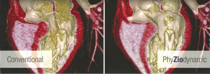
The system takes CT or MR data sets of a full cardiac cycle, allows manipulation to create a 3D rendering of the anatomy of interest, and then puts the data set in motion in a video loop. The resulting video image is similar to the functional movement seen in angiography cine loops, but the color and more detailed anatomy greatly enhances the image and the information it can offer.
The company displayed examples of the high-definition, animations showing the functioning of an aortic valve and a cut-away of an entire heart. Some cardiac surgeons who saw the examples said it was the first time they had seen such detailed functional of the anatomy outside of live, open surgery. The cut-away of the heart clearly showed the movement of the papillary muscle and the chordae tendinae.
The initial images appear a lot like live, color, 3D echo, where there is a lot of noise and artifacts. The software helps filter the majority of this noise to create a much clearer image.
The software solutions are based on supercomputing technology originally developed for dynamic, instantaneous weather pattern analysis. Over the years, supercomputing has evolved from a large room of host computer hardware to a centralized, single CPU-based processing server allowing multiple concurrent users throughout the enterprise system. This has resulted in several significant advantages including superb image quality, optimized user efficiency and maximum cost effectiveness for an extensive range of clinical applications.
The technology offers scalable software solutions, eliminating the need for costly, multiple proprietary or specialized GPU rendering boards. It integrates seamlessly with standard, off-the-shelf hardware and expands the life of expensive modalities such as CT and MR.
Through the application of advanced algorithmic processes, the technology reduces noise without compromising image integrity, allowing the physician to increase diagnostic confidence. It also handles extremely large sets of data on an enterprise basis, rendering images and analysis automatically for greater patient throughput without the need for proprietary or specialized GPU rendering hardware. Ziosoft said the key to its technology is the use of uncompressed, lossless images, which ensure the integrity of all the data. The images are accessible from the office, home or enterprise without the need for proprietary hardware. PhyZiodynamic technology is compatible with any DICOM imaging modality including CT, MR, PET, SPECT and angio. This allows advanced diagnostic applications for multiple medical specialties such as cardiology, radiology, neurology, orthopedics and oncology. Ziosoft has received regulatory clearance for several applications including CT coronary analysis and calcium scoring, as well as both MR and CT cardiac function analysis.
Ziosoft hopes to create a commercial version of the 5D analysis software in the next 12 months.
For further information: www.ziosoftinc.com

 April 18, 2025