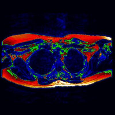
Pictured is a digitally enhanced axial MRI of the upper chest (as if viewed from the feet). Areas of potential brown fat are shown in green. Courtesy University of Warwick, UHCW.
April 18, 2014 — The first MRI (magnetic resonance imaging) scan to show 'brown fat' in a living adult could prove to be an essential step toward a new wave of therapies to aid the fight against diabetes and obesity.
Researchers from Warwick Medical School and University Hospitals Coventry and Warwickshire NHS Trust have used an MRI-based method to identify and confirm the presence of brown adipose tissue in a living adult.
Brown fat has become a hot topic for scientists due its ability to use energy and burn calories, helping to keep weight in check. Understanding the brown fat tissue and how it can be used to such ends is of growing interest in the search to help people suffering from obesity or are at a high risk of developing diabetes.
Dr. Thomas Barber, department of metabolic and vascular health, Warwick Medical School, Coventry, England, explained, "This is an exciting area of study that requires further research and discovery. The potential is there for us to develop safe and effective ways of activating this brown fat to promote weight loss and increase energy expenditure – but we need more data to be able to get to that point. This particular proof of concept is key, as it allows us to pursue MRI techniques in future assessments and gather this required information."
The study, published in the Journal of Clinical Endocrinology and Metabolism, outlines the benefits of using MRI scans over the existing method of positron emission tomography (PET). Whilst PET does show brown fat activity, it is subject to a number of limitations, including the challenge of signal variability from a changing environmental temperature.
Unlike PET data which only displays activity, MRIs can show brown fat content whether active or not – providing a detailed insight into where it can be found in the adult body. This information could prove vital in the creation of future therapies that seek to activate deposits of brown fat.
"The MRI allows us to distinguish between the brown fat and the more well-known white fat that people associate with weight gain, due to the different water to fat ratio of the two tissue types. We can use the scans to highlight what we term 'regions of interest' that can help us to build a picture of where the brown fat is located,” added Barber.
Barber works at the Human Metabolism Research Unit at UHCW. The unit has benefitted from substantial investment through the Science City Research Alliance, a large-scale, long-term research program between the University of Birmingham and the University of Warwick.
For more information: www.warwick.ac.uk

 October 01, 2026