June 12, 2015 - A new molecular imaging agent has been demonstrated to find prostate cancer that has spread to other tissues by locking in on the prostate specific membrane antigen (PSMA) enzyme, according to a new study. Results were presented at the 2015 Annual Meeting of the Society of Nuclear Medicine and Molecular Imaging (SNMMI).
"To date, conventional imaging is limited in detecting prostate cancer metastasis accurately and measurably," said Neeta Pandit-Taskar, M.D., co-author of the study and a researcher at Memorial Sloan Kettering Cancer Center in New York City. "Using this agent, we can detect the prostate cancer cells that have metastasized to bone - one of the most difficult areas to evaluate using standard methods. We hope that this research will help us develop earlier and more effective detection of disease and assist in clinical decision-making."
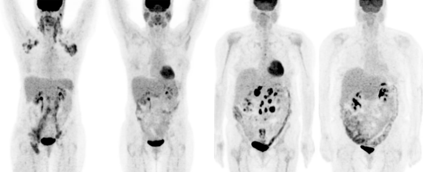
The radiotracer combines a small amount of the radioactive material zirconium-89 with a fragment of an antibody called a minibody. This minibody has anti-PSMA qualities and attaches to overexpression of the enzyme (more technically known as glutamate carboxypeptidase II or GCPII) on the exterior of prostate cancer cells wherever they may have traveled in the body. The novel radiotracer, called Zr-89 Df-IAB2M (IAB2M), is imaged faster than the full antibody (J591) that targets PSMA and has been shown to be safe for patients. Particles emitted from the site are then detected by positron emission tomography (PET), which creates a computerized image of the activity of the agent within the body. The resulting scan highlights "hot spots" of PSMA overexpression.
"Initial results of full-body imaging with this Zr-89 radiolabeled minibody have shown that we are able to detect more disease sites in patients than with conventional imaging," said Pandit-Taskar. "With further validation, this radiotracer could also potentially be used to perform targeted biopsies for precise tissue analysis, which could lead to earlier, more appropriate treatment for prostate cancer patients."
For this research, a total of 28 subjects were imaged with a variety of imaging modalities, including standard imaging with computed tomography (CT), magnetic resonance imaging and molecular bone scan (SI); PET with a common radiotracer called fluorodeoxyglucose (FDG-PET); and PET with CT and the agent Zr-89 IAB2M (IAB2M PET/CT) assessed in escalated doses. A selection of suspected disease sites were then biopsied.
Results of the study showed there were a total of 393 suspected lesions found in soft tissue and bone using all imaging modalities, collectively. IAB2M PET/CT identified 81.7 percent of all suspected bone lesions, and 65 of these growths that were not identified with any other modalities. Additionally, IAB2M found 32 soft tissue lesions not found by other methods.
Of the total 19 biopsies and histological analyses performed, FDG-PET identified 14 growths, IAB2M PET/CT identified 17 growths, and SI found 18. However, overall accuracy of positive/negative reading was found to be 89.5 percent for IAB2M PET/CT versus 84 percent for SI and 84 percent for FDG-PET.
"This is an early phase trial," said Pandit-Taskar. "For the next few years, we will be exploring IAB2M in additional clinical trials. More data are needed to understand its clinical value but, if results are favorable, this imaging agent could play a critical role in the standard of care for prostate cancer."
An estimated 220,800 new cases of prostate cancer and 27,540 prostate-cancer related deaths occur annually in the U.S. alone. As many as one out of every seven American men are expected to be diagnosed with prostate cancer at some point in their lifetime, according to 2015 statistics from the American Cancer Society.
For more information: www.snmmi.org

 July 15, 2026


