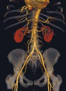
The level of detail CT scans provide, such as those produced by the Aquilion 64, can lead to faster, more accurate diagnoses.
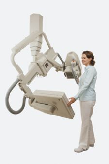
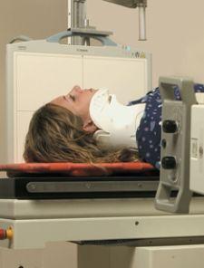
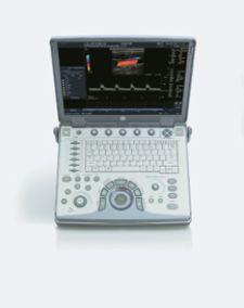
Today’s busy emergency departments have an arsenal of imaging tools at their disposal when triaging and diagnosing trauma patients. Increasingly, ED physicians are turning to more sophisticated technologies in addition to proven standbys — such as ultrasound, CR and DR — when diagnosing victims. Modalities can vary from hospital to hospital and are determined by numerous factors, including cost, availability and injury type. Making Waves Ultrasound has long been a staple in the ED and its importance is steadily growing. At Denver Health Medical Center, ultrasound can have a significant impact on patient health, says John L. Kendall, M.D., FACEP. Ten years ago, according to Dr. Kendall, ultrasound in the ED was used primarily for trauma. Today, its indications in emergency medicine have grown considerably and include pericardial infusion, abdominal aortic aneurysm, ectopic pregnancies, gallbladder disease, renal stones and more. Denver Health’s ED also uses ultrasound a great deal for image-guided procedures. In the ED, where time can be a physician’s greatest enemy, the ability to expedite patient care is a huge benefit. “Ultrasound’s advantages are not limited to just accuracy of diagnosis, but also include speed of disposition, and studies have shown it can improve patient care from the standpoint of mortality as well,” Dr. Kendall explained. “We found that the length of stay from the time that the patient is in the emergency department can be decreased by about two hours if we are using the ultrasound as opposed to ordering the study from the radiology department.” Last year, Denver Health’s ED was approached by GE Healthcare to clinically evaluate the company’s latest portable ultrasound unit, the LOGIQe, which is designed for quick, accurate decision-making. Up until the evaluation began, the department had never housed a GE machine. For three weeks, the busy ED put the unit through its paces and was very impressed with how the machine responded. First of all, “it imaged fantastically right out of the box,” Dr. Kendall said. “We found the LOGIQe easy to use, and it quickly became the machine of choice within the department,” he added. Additional features such as report generation and the unit’s potential ability to embed images within a report make it a great fit for Denver Health’s ED needs, Dr. Kendall says. While certainly not replacements for full-sized ultrasound equipment, portable units definitely have found a niche in the ED. SonoSite, maker of a comprehensive line of portable ultrasound devices, is dedicated to ensuring users make the most of the technology’s diagnostic potential and procedural applications throughout the hospital. In June 2006, the company launched an initiative that will allow clinicians to view refresher courses via Apple’s fifth-generation iPod with video. “Ultrasound is dynamic, ‘real-time’ technology,” said Daniel D. Price, M.D., staff emergency physician at Highland General Hospital, Oakland, CA, and director of the Emergency Ultrasound Fellowship. “Focused, well-designed one-to-five-minute tutorials can be viewed on an iPod wherever, whenever they are needed, and can bring the physician or medical student up to date, on the spot,” he explained. Time is Life When diagnosing trauma patients, speed combined with accuracy is paramount. Both computed radiography (CR) and digital radiography (DR) expedite triage in the ED by providing quality digital X-ray images quicker than film-based X-ray systems and with less radiation than CT or PET. CR systems currently boast a lower cost and are easy to use; yet, some believe DR may eclipse CR as the preferred digital X-ray option, especially now that it is portable. “Emergency rooms today are overwhelmed with patients, so you have to handle more patients in less time,” said Helen Titus, DR Product Line manager, Kodak’s Health Group. “With an image every 20 seconds, you can see the workflow advantage of the DR. In addition, DR offers superior image quality,” Based on what staff radiologists at Strong Memorial Hospital are reporting, Mary Ellen Wilson, chief radiologic technologist and Kathy Orsini, emergency department radiology supervisor, agree. The Level 1 trauma center at the Rochester, NY hospital is also experiencing other DR benefits, such as lower dose and fewer retakes. But what makes the biggest difference both in terms of workflow and patient care, says Orsini, is that there is no stopping to change cassettes; the less you have to lift or move a trauma patient, and the quicker you can expedite their care, the better, she says. The Imaging Science department. in Strong’s ED currently has six DR units: four Kodak DirectView DR 7500s and two DirectView DR 9000s. Thanks to the same operator interface on all Kodak CR and DR units, the learning curve for techs is about two weeks, and the transition between working with CR and DR systems is virtually seamless. Since integrating DR into its ED workflow about a year and a half ago, Overlake Hospital Medical Center (Bellevue, WA) has seen an immense increase in exam speed, says Kay Daughtry, radiology supervisor. “The minute you take your exposure the image shows up on the computer screen,” explained Daughtry. And because prior images are immediately erased, it enables the techs to do a sequence of X-rays quickly, she says. Overlake uses Canon’s CXDI-50G portable DR unit as well as the stationary CXDI-40G, which are both housed within the X-ray department located directly adjacent to the ED. The Canon DR room, as the staff calls it, is definitely the favored room for techs, says Daughtry. “The techs will wait to use that room,” Daughtry said, which was sold and installed by Canon dealer Core Medical in Kenmore, WA. Daughtry estimates that 80 percent of their work is performed there, primarily because (1.) the systems are easy to use and (2.) necessary images can be obtained quickly. Images can also be manipulated; techs can focus in one area and can change density and contrast. In addition, once the exam is closed out, images are immediately available for physician review. Initially concerned as to how the CXDI-50G would hold up to the department’s workload, Daughtry says the unit is quite durable, and they are very pleased with its performance. So pleased, in fact, that for their new ED, which is presently under construction, they are adding another Canon DR room that will be basically identical to its predecessor. Diagnosis is in the Details Revered for its ability to present anatomical information in precise detail, CT has become a diagnostic favorite among physicians. The systems can generate eight, 16 and 64 slices of data in a single breathhold. In fact, multislice CT scans can locate injuries from head to toe and provide images of most everything in between, including the vasculature and heart function. With all CT’s advantages, is any other modality even needed for trauma imaging? Maybe not, according to Shahid Shafi, M.D., assistant professor of Trauma at Parkland Memorial Hospital, University of Texas Southwestern Medical Center, Dallas. Dr. Shafi and fellow presenters Drs. Amy Sisley and Heidi Frankel shared their opinions regarding the role of CT imaging in treating trauma injuries at the 2006 Society of Critical Care Medicine’s annual congress, reporting that CT is far more sensitive than traditional chest X-ray in identifying most common injuries that result from blunt traumas. In the case of penetrating trauma, such as a gunshot wound, CT can identify the bullet’s missile tract, essentially guiding the physician directly to the injury. Because of the definition and the level of detail it provides, CT can also help physicians select patients for nonoperative management of solid organ injuries. The news is also positive regarding CT neuro trauma scanning and angiography. For neurological injuries alone, according to Dr. Frankel, additional imaging is probably not necessary, and she added, carotid CTA is “a good enough study” to exclude blunt carotid and vertebral artery injury. When it comes to recognizing the benefits of CT scans for trauma patients, Frederick M. Barnett, M.D. needs no convincing. As director of Radiology Services at Metroplex Hospital, Kileen, TX, Dr. Barnett sees the level of detail CT scans provide every day, detail which he says leads to faster, more accurate diagnoses and better patient care. The community hospital treats 45,000 ED cases per year, covering all types of trauma injuries including neurological, musculoskeletal, gunshot wounds and motor vehicle accidents. “Our protocol is quite simple,” said Dr. Barnett. “The multiple trauma patient is scanned with a 64-slice CT (Toshiba’s Aquilion 64) from which the images can be quickly reconstructed and reformatted so they can then be viewed in any plane or in 3-D to accommodate the ED physician’s needs.” CT’s ability to image, for example, the chest, abdomen and pelvis in 15-20 seconds often obviates the need for sedation — a huge benefit, especially for pediatric patients and those who may be experiencing altered mental status, says Dr. Barnett. However, the most compelling benefit of multidetector CT (MDCT) scanning, he stresses, is that it eliminates the steps previously required in multiple trauma cases — and the delay in treatment that can result — from first having to rule out spinal injury. Because MDCT systems produce thin-slice isotropic, volumetric data sets, the radiologist can manipulate CT scan data on a workstation to produce sagittal, coronal and oblique multiplanar reformations of spinal images from an initial, single CT scan. This, Dr. Barnett explained, is key to expediting accurate treatment. The reformatted CT datasets are as good or better than plain films in clearing the spine for fracture. The CT scan is a rapid, comprehensive and noninvasive tool that can be ideal for imaging unstable trauma patients; however, as Dr. Shafi recommends — and this is good advice for all modalities — when imaging trauma victims, physicians should be familiar not only with the benefits, but also the possible risks, limitations and alternatives.


 July 10, 2026


