The explosion in diagnostic imaging studies in emergency departments (EDs) has led to the need for radiologists to interpret examinations 24 hours a day, seven days a week.
Imaging in the ED continues to grow as doctors increasingly rely on medical imaging to triage patients. Magnetic resonance imaging (MRI), computed tomography (CT) and positron emission tomography (PET) scans accounted for 14 percent of all ER visits in 2007, according to the Centers for Disease Control and Prevention.
With a large portion of the U.S. population reaching 65 years and older by 2020, diagnostic imaging is predicted to continue its upward spiral. As the frontline of care relies increasingly on imaging, the speed and accuracy of the technology becomes more critical. This is especially important for patients who present with potentially life-threatening conditions, such as stroke and chest pain.
DR Demand Grows
The majority of diagnostic imaging tests conducted in the ED are radiographic (X-ray) images. X-rays, whether acquired on a computed radiography (CR) or digital radiography (DR) system, made up 35 percent of all diagnostic imaging in 2006.1 These systems must be portable for patient comfort, flexible enough to capture images at all angles of the anatomy and able to quickly develop images to facilitate fast and accurate diagnosis.
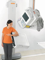
New wireless DR systems are in increasing demand, as the untethered detectors are well-suited for producing digital images in confined spaces, emergency departments, sterile environments and the intensive care unit (ICU), as well as for positioning around patients with limited mobility and during remote imaging.
Patients undergoing X-ray exams often suffer from painful debilitation where mobility is limited. They may be using a wheelchair or are unable to stand or sit up. Wireless detectors make it easier for technologists to capture images from hard-to-reach angles.
The first company to release a wireless DR cassette in the U.S. market was Carestream Health with the DRX-1 system. The standard cassette size is designed to fit existing X-ray rooms without having to replace or modify existing equipment.
Many digital radiography manufacturers now offer wireless cassettes because of the flexibility it offers in imaging. Another spin on DR cassettes is to provide a detachable power supply. The FDR D-EVO by Fujifilm Medical Systems USA gives users the option to untether the cord or hook it up to the system to charge the battery.
Clinicians at the Dartmouth-Hitchcock Medical Center (DHMC), New Hampshire, emergency department are using the recently released Philips’DigitalDiagnost X-ray system with a wireless detector.
“You have a digital cassette that is completely untethered and can be placed in virtually any position,” said Jim Roberts, administrative director of radiology at DHMC. “We are currently taking advantage of this capability in the emergency department where we find it provides many of the same benefits as in the typical ICU portable setting. We can use it on all trauma patients. If we need to keep a patient on a stretcher, we can place the wireless detector under the patient. If we [are dealing with] multiple trauma, a rollover in a vehicle or someone who is very unstable, then we have versatility.”
The wireless DR detectors directly compete with portable computed radiography (CR) units.
“We are absolutely confident we have been able to reduce the patient stay by using this detector, because we don’t have to move the patients. CR cassette usage has dropped to almost none. We know we have shaved minutes off of the patients’ stay,” Roberts added.
Recently, IMIX Americas Inc. integrated a wireless DR cassette and software as part of its IMIX DR system.
Speed is another advantage of DR systems. Konica Minolta Medical Imaging introduced the Xpress DR, which provides image previews in about five seconds and touts the ability to take exposures every 10 seconds. Carestream’s DRX-1 system delivers preview images in about five seconds, and D-EVO transmits images to the technologist workstation in five seconds and boasts nine-second cycle times.
“With the DigitalDiagnost, you can send the image directly to your picture archive and communication system (PACS) or archive. So speed is definitely the advantage. It takes about six seconds to develop an image,” Roberts noted. “The biggest benefit has been patient comfort and speed.”
Today, Roberts will carry the detector no more than 25 feet from the X-ray room. But he believes the future of this technology lies in allowing the clinicians to carry the detector anywhere around the hospital.
Door-to-Brain Time
From the moment the patient is wheeled into the emergency department to the time the patient is diagnosed and treated, the rapid-response protocol for critically ill stroke patients developed by the Mayo Clinic in Rochester, Minn., includes a CT scan within 10 minutes of arrival at the local emergency room. This serves to determine the type of stroke — ischemic or hemorrhagic — and allow treatment to begin.
A thrombolytic medication is injected intravenously to dissolve the clot for patients whose stroke is ischemic and whose symptoms began less than three hours before arrival at the hospital. The thrombolytic medication increases the chances of a full or almost-full recovery by one-third.
With strokes, time equals brain cells. So the need to immediately diagnose the type of stroke and extent of the damage is critical.
Typically, for stroke workup, one of the first exams the emergency department will do is a noncontrast CT of the head to rule out the hemorrhage. About 75 to 80 percent of the time, there is no hemorrhage. If there is no hemorrhage, the next step is to do a CT perfusion to determine how much of the brain is deprived of oxygen. One of the fastest tools for analyzing stroke is the Aquilion One by Toshiba America Medical Systems. The 320-detector row CT system can capture the entire brain in one rotation.
“The problem is, you don’t know where in the brain the hemorrhage is. That is why you want to look at the entire brain,” said Rich Mather, senior manager of clinical programs, Toshiba’s research department.
“Conventional technology can only cover about 4 cm of the head, which is about a quarter of the brain in the dynamic imaging,” Mather said. “That only allows you to look at where the most likely places are that a stroke may have occurred, but you run the risk of missing anatomy and missing the location of the stroke.
“With the Aquilion One’s 16 cm of coverage, you get the whole brain,” he continues. “This allows you to do whole-brain perfusion and find out where that stroke is. It is a very rapid and accurate method of looking at dynamic perfusion in stroke patients, and it is great for the emergency department setting.”
Some of the centers that use the Aquilion One routinely have optimized their workflow, reducing the time it takes to do the whole procedure from the time they press the button on the scanner until the time they are reading the perfusion mounts on the console, to about four minutes.
Treating Chest Pain
Chest pain is one of the most common symptoms of patients presenting to an emergency department. In the United States, more than 5 million patients a year come to the emergency room with a chief complaint of chest pain.
Emergency department use of 256-slice computed tomography angiography (CTA) can help physicians triage patients with indeterminate chest pain without the need for additional diagnostic testing.
“Traditional evaluation of chest pain in the emergency department is frequently inconclusive and often requires the admission of patients for further diagnostic testing, which is costly,” said Minh Lu, M.D., nuclear medicine resident, department of radiology and nuclear medicine, University of Maryland Medical Center, Baltimore, Md. Lu is the author of a study2 performed at the university, in which 11 patients underwent a CT angiography (CTA) exam on a 256-slice CT system. Doctors evaluated their indeterminate chest pain.
Seven patients were found to have a negative CTA and a final clinical diagnosis of insignificant chest pain. Two of the patients had insignificant coronary plaque. Two others had moderate coronary disease, but were given presumptive final diagnoses of noncardiac chest pain. In addition, there were two pulmonary findings and one breast mass found incidentally.
“Overall, the diagnostic concordance of 256-slice CTA was 100 percent,” Lu said. “Traditional management of chest pain may require observation prior to a radionuclide stress study or stress echocardiogram before discharge, increasing the length of hospital stay and cost. In contrast, 256-slice cardiac CTA can be performed safely and early in the observation period with rapid and accurate results.
“The 256-slice cardiac CTA shows substantial promise in expediting and improving emergency department triaging of patients presenting with chest pain,” Lu adds.
Physician reliance on diagnostic imaging to diagnose patients in the ED is destined to grow exponentially. As imaging modalities enhance their speed and accuracy, clinical utility will drive their further adoption.
Reference:
1. National Health Statistics Reports. Number 7. August 6, 2008.
2. Minh, L., et al. “Ultra Low Dose MDCT Evaluation of the Axial Skeleton: A Comparison of Dose and Subjective Clinical Efficacy.” American Journal of Roentgenology. 2010; 194:A1-A4.


 June 24, 2026


