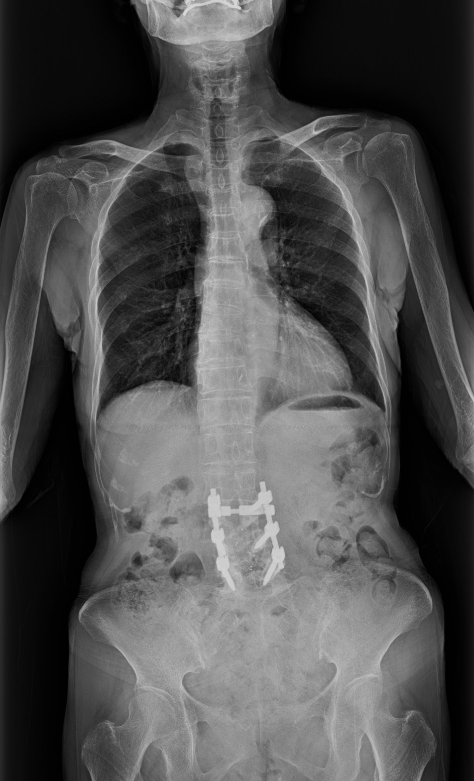
Advances in long-length digital radiography are creating opportunities for visualization during spinal surgery, as well as pre- and post-operatively. Image courtesy of Fujifilm Medical Systems
Recent advances in digital radiography (DR) are increasing demand for whole spine imaging before, during and after back surgery, according to Gregg R. Cretella, national director of clinical operations for Fujifilm Medical Systems, USA.
“Spine surgeries in the United States — probably worldwide — are on the rise,” he told Imaging Technology News. “Digital radiography vendors have (developed) advanced DR technology that better supports spine imaging.”
At the Association for Medical Imaging Management (AHRA 2019) meeting on July 24, Cretella will explain how DR long-length detector technology and image processing algorithms are changing the utilization of this modality, specifically in spine imaging. He plans to describe how these advances can help improve patient comfort, reduce radiation dose, and drive clinical, operational and financial outcomes.
Discussions will address DR as well as computed radiography (CR) for whole spine and other applications of long-length imaging. Because CR cassettes are still in widespread use, this modality continues to figure into spine imaging, just as different DR methods can be used for these applications, according to Cretella.
Long-length Imaging Methods
CR, when used for long-length imaging, may require manually setting up cassettes and then stitching the resulting images together. Similarly, if done with a small detector, digital radiographs may also have to be stitched together. The process can be grueling. But semiautomatic and automatic methods are available.
The ultimate solution is to use a single long-length DR detector that produces an image of the entire spine. “The biggest advantage (of the long-length detector) is that it allows for a single exposure of the entire region of clinical interests,” Cretella said. “And, because it is digital radiography technology, there is almost immediate gratification from that single exposure,” noting that surgeons can look at the image soon after the exposure.
The use of long-length detectors may also improve patient comfort, he said. The reason is the fast data acquisition, which may be especially appealing to patients during the post-operative phase, Cretella opined: “After back surgery, I would imagine that standing is a challenge.”
Patient safety may also be improved through the use of DR, he said. Reductions in radiation dose from 50 to 75 percent may be achieved using DR long-length technologies compared with those of CR, according to Cretella. He plans to address advances in dose monitoring during his talk, as well as “exposure surveillance” specific to whole spine imaging.
“Ultimately you will be able to use these tools to continue to reduce patient dose for whole spine imaging,” he said, “and then to facilitate any dose reduction programs that you may have.”
Cretella said he hopes the information he presents at AHRA will help administrators considering the acquisition of long-length imaging technologies, particularly in light of shifting utilization. Until the last year or so, this type of radiography was focused primarily on scoliosis and long-leg imaging outside the operating room. Recently, however, surgeons have been demanding whole spine images during the operative procedures, he said.
Greg Freiherr is a contributing editor to Imaging Technology News. Over the past three decades, Freiherr has served as business and technology editor for publications in medical imaging, as well as consulted for vendors, professional organizations, academia, and financial institutions.
Editor’s note: This article is the fourth piece in a content series by Greg Freiherr covering The Association for Medical Imaging Management (AHRA) annual meeting in Denver. The first article, How Standardizing Protocols Can Save Time and Money, can be found here. The second article, How Artificial Intelligence Might Impact Radiology, can be found here. The third article, How To Manage Risk in the MR Suite, can be found here.
Related content:
How Standardizing Protocols Can Save Time and Money
How Artificial Intelligence Might Impact Radiology
How To Manage Risk in the MR Suite
Fujifilm Unveils Long-Length Digital Radiography Detector at RSNA 2015
Scoliosis Imaging: What Radiologists Should Know


 July 23, 2026


