
Technology
November 7, 2011 — iCAD Inc. will highlight a new, works-in-progress magnetic resonance image-guided (MRI) prostate biopsy prototype. The system features PrecisionPoint for VividLook, a combination probe and positioning software. It uses in-plane scan specification and localizer scans to determine position of the needle, and a semi-rigid arm that allows for a broader range of motion.
 November 07, 2011
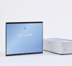
Technology
November 7, 2011 — Konica Minolta’s new portable X-ray upgrade kit efficiently turns portable X-ray systems into digital, wireless solutions. The Aero DR Portable Retrofit Solution is designed for a quick, easy and inexpensive transformation from analog to digital. With a very small footprint, the unit can be installed and stored inside the cassette storage bin.
November 07, 2011 
Technology
November 7, 2011 — The InfiniVault from Rorke Data Inc., an Avent company, sets a new standard in enterprise archive storage for healthcare providers. Its data archive appliances are specifically designed for mid-size enterprises and small healthcare facilities that have rapid, unpredictable growth in unstructured data and rigorous data retention requirements.
November 07, 2011 Sponsored Content
Sponsored Content
Videos | Radiology Imaging
Today’s radiology teams are faced with a wide range of growing complexities such as patient and procedure variabilities ...
June 24, 2026 
Technology
November 7, 2011 — Sectra’s Web-based picture archiving and communication system (PACS) is optimized for wide area radiology, enabling efficient workload sharing across multiple sites. The patented technology, Sectra RapidConnect, allows for efficient transfer even of large stacks over strained networks. The system features a vendor-neutral sharing solution based on XDS-I for immediate access to all clinical data from any connected discipline, independent of location.
November 07, 2011 
Technology
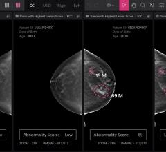
November 7, 2011 — Sectra Breast Imaging PACS (picture archiving and communication system) streamlines workflows of high-volume mammography screening and advanced diagnostic breast imaging procedures. It features true multimodality capabilities and now supports display and review of breast tomosynthesis images.
November 07, 2011 
News
November 7, 2011 — This year’s annual meeting of the European Association of Echocardiography (EAE), a registered branch of the European Society of Cardiology (ESC), is changing its name to “EUROECHO & other Imaging Modalities.”
November 07, 2011 Sponsored Content

Feature | Information Technology
AT A GLANCE Organization: Expert Radiology Management Services, LLC Specialty: Subspecialty teleradiology — neuro and ...
May 01, 2026 
News
November 7, 2011 – The Society of Nuclear Medicine (SNM) launched a new patient-focused website, discoverMI.org, to provide patients with information about nuclear medicine and molecular imaging and how it can play a critical role in the detection, treatment and management of diseases.
November 07, 2011 
News
November 7, 2011 -- Philips combines its Ambient Experience with Gemini TF Big Bore positron emission tomography (PET)/computed tomography (CT) at Sparks Regional Medical Center, Arkansas’ first hospital, serving a population of more than 350,000 in and around its location in Fort Smith.
November 07, 2011 News
November 7, 2011 – A California jury recently found Hologic guilty of violating two patents in a case filed by Conceptus Inc.
November 07, 2011 Sponsored Content
Sponsored Content
Videos | Radiology Business
Radiology departments have many different needs and face a wide variety of challenges that can impact their departments ...
November 11, 2025 
Technology
Cerner cardiovascular solutions enable cardiologists to automate functions across the continuum of care. Built upon the Cerner Millennium architecture, Cerner’s PowerChart Cardiovascular solution unifies cardiovascular diagnostic activities, therapeutic interventions and follow-up regimens into a single workflow, integrated with the electronic health record (EHR). PowerChart Cardiovascular gives the cardiologist the ability to view images in context with Cerner’s cardiovascular workflow manager utilizing the integration with the Merge viewer.
November 03, 2011 
Technology
New tools for Brit Systems’ WebWorks pure browser-based viewer and DoctorWorks iPad viewer help clinicians access images anywhere, anytime. BRIT's development team strives to enhance a physician's lifestyle with access to images from any Internet browser or iPad application. The new WebWorks tools include: Cine cardiac ultrasounds and optional MPR capabilities; and transferring measurements from the image viewer directly into report forms. DoctorWorks 1.1 for the iPad features the simultaneous viewing of two studies and the addition of a timeline of studies on both the study information screen and the viewing screen. DoctorWorks is available from the iTunes App Store.
November 03, 2011 Technology
CareAware MultiMedia is Cerner’s vendo-neutral archive (VNA) platform allows management of multimedia, such as images and video and integration with the electronic health records (HER). This ensures that important information is contextually relevant, comprehensive and timely — an important aspect of any organization's imaging strategy. Critical information no longer resides in isolated, disconnected silos. It is available at the point-of-care, or wherever clinicians need it.
November 03, 2013 Sponsored Content
Feature | Breast Imaging
Despite decades of progress in breast imaging, one challenge continues to test even the most skilled radiologists ...
October 24, 2025 
Case Study | Intelerad Medical Systems
Intelerad’s InteleOne distributed radiology solution enables a regional healthcare provider to reduce costs while maintaining and improving its focus on accessibility, convenience and high levels of quality and service.
November 03, 2011 
News
November 3, 2011 – Lantheus Medical Imaging announced important changes to the U.S. product label for Definity Vial for (perflutren lipid microsphere) Injectable Suspension. The contrast agent is indicated for use in patients with suboptimal echocardiograms to opacify the left ventricular chamber and to improve the delineation of the left ventricular endocardial border.
November 03, 2011 
Technology
Cerner and Vital Images bring care providers advanced visualization across the health care continuum. The system provides workflow-driven multimodality 2-D, 3-D and 4-D viewing tools to enhance decision support in context with patient information via the electronic health records (EHR) for real time analysis and decision support.
November 02, 2011 
Technology
The Cypher PS print server and Canon imagePress C1+ digital printing solution is a cost-effective answer that enables radiology departments to print high-quality digital imaging and communications in medicine (DICOM) images on various types and sizes of paper. This digital printing solution helps to cut the cost of expensive film usage by providing a low-cost, high-quality alternative.
November 02, 2011 
Technology
MediCal QAWeb is the industry’s only completely online, automated quality assurance (QA) and auto-calibration service that guarantees maximum diagnostic confidence and uptime of all picture archive and communications system (PACS) display systems throughout the healthcare facility. This all-inclusive, intervention-free service ensures 24/7 digital imaging and communications in medicine (DICOM) compliance for every medical imaging display on the network and maintains consistent luminance to enhance diagnostic confidence. Offering centralized control of all displays across the enterprise, Barco’s MediCal QAWeb provides fast issue identification and automated corrective actions to ensure maximum availability. Asset management capabilities enhance budget control with comprehensive display analysis and reporting. For more information: www.barco.com
November 02, 2011 
Technology
Barco's Mammo Tomosynthesis 5 MP significantly improves accuracy of breast cancer detection with numerous capabilities that enhance the conspicuity of the subtlest details. RapidFrame technology counteracts motion blur when scrolling through a stack of images due to a high pixel refresh rate. images and eliminates quantization artifacts, reducing the overall noise in the images.
November 02, 2011 
Technology
Invivo’s DynaCAD for Prostate and DynaTRIM are an award-winning combination (Medical Design Excellence Gold Award, 2010). The software and hardware combination streamlines workflow and improves productivity and increases diagnostic confidence for both imaging and magnetic resonance (MR)-guided intervention.
November 02, 2011 
Technology
Gamma Medica Inc. will demonstrate the LumaGEM Low Dose Molecular Breast Imaging (MBI) System at RSNA 2012.
November 02, 2011 © Copyright Wainscot Media. All Rights Reserved.
Subscribe Now