
News
Barco, a visualization technology company, and Brainlab AG, Germany-based medical software technology manufacturer and marketer, announced their collaboration to provide medical content distribution and sharing.
 January 06, 2014
 Blog
“Those who cannot remember the past are condemned to repeat it.” —George Santayana, 20th century philosopher and writer
January 06, 2014 
Feature
University of California, Davis radiologists, medical physicists and orthopaedic surgeons have found a way to create "movies" of the wrist in motion using a series of brief magnetic resonance imaging (MRI) scans.
January 06, 2014 Sponsored Content
Sponsored Content
Videos | Radiology Imaging
Today’s radiology teams are faced with a wide range of growing complexities such as patient and procedure variabilities ...
June 24, 2026 
News
GE Healthcare showcased its SensorySuite “cocoon” experience at the Radiological Society of North America Annual Meeting (RSNA 2013).
January 06, 2014 News
Novarad ended 2013 closing contracts and is finalizing installations for nine facilities.
January 06, 2014 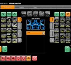
Technology
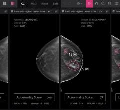
Ikonopedia announced the launch of its SonoPro breast ultrasound interpretation and reporting module, designed to guide radiologists through the interpretive process for diagnostic breast ultrasound exams.
January 03, 2014 Sponsored Content

Feature | Information Technology
AT A GLANCE Organization: Expert Radiology Management Services, LLC Specialty: Subspecialty teleradiology — neuro and ...
May 01, 2026 
Technology
GE Healthcare MRI announced DV24.0, a software platform featuring applications such as Silent Scan.
January 03, 2014 News
The American Society of Radiologic Technologists (ASRT) has launched its new educational series, Image-guided Radiation Therapy.
January 03, 2014 
News
As the global medical isotope shortage continues indefinitely following the plant leak in South Africa, Berks Cardiologists Ltd., a cardiovascular imaging center in central Pennsylvania, is continuing to operate at the same procedure volume and with their lower-dose cardiac nuclear medicine imaging protocols.
January 03, 2014 Sponsored Content
Sponsored Content
Videos | Radiology Business
Radiology departments have many different needs and face a wide variety of challenges that can impact their departments ...
November 11, 2025 News
Imaging research from Western University, London, Canada, has demonstrated that a magnetic resonance imaging (MRI) approach called quantitative susceptibility mapping (QSM) can be an important tool for diagnosing and tracking the progression of multiple sclerosis (MS) and other neurological diseases.
January 02, 2014 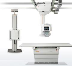
News
Carestream Health has been awarded an extension of its contract with the U.S. Department of Defense that allows the federal government and federal agencies to spend up to $70.2 million for digital radiography (DR) and computed radiography (CR) medical imaging systems.
January 02, 2014 
Feature
The American College of Radiology (ACR) supports the United States Preventive Services Task Force (USPSTF) recommendation (Grade B) for low-dose computed tomography (CT) lung cancer screening of adults 55 to 80 years old who have a 30 pack-year smoking history and currently smoke or have quit within the past 15 years.
January 02, 2014 Sponsored Content
Feature | Breast Imaging
Despite decades of progress in breast imaging, one challenge continues to test even the most skilled radiologists ...
October 24, 2025 Technology
Focal Therapeutics announced that its BioZorb 3-D tissue marker has been successfully used in over 100 patients.
January 02, 2014 
News
Fujifilm Medical Systems U.S.A. Inc., offered the latest version of Synapse RIS (radiology information system), a complete electronic health record (EHR)-certified solution that can be adapted to meet Meaningful Use (MU) Stage II requirements for radiology professionals.
January 02, 2014 
Technology
Fujifilm Medical Systems U.S.A. Inc., featured its newest women’s health products including the Aspire Comfort Paddle, a patented paddle designed to improve patient comfort, at the Radiological Society of North America Annual Meeting (RSNA 2013) in Chicago.
January 02, 2014 News
GE Healthcare and Konica Minolta Inc. have signed a global distribution agreement of Konica Minolta’s AeroDR cassette-size digital X-ray imaging retrofit. Having built a collaborative relationship for nearly ten years related to the sales of computed radiography in the United States, the two partners have now agreed to expand their strategic alliance, with GE Healthcare distributing AeroDR not just within the United States, but globally as well, using GE Healthcare sales channels.
January 02, 2014 
Technology
Accelarad, a cloud-based medical image sharing solution, announced a product update at the Radiological Society of North America Annual Meeting (RSNA 2013) that includes image exchange analytics reporting and a new user interface (UI).
January 02, 2014 News
Massachusetts was on the same brink in 2006 that the entire nation is on today: the brink of expanding health insurance to cover far more people than before through government-driven, market-based reform.
December 31, 2013 News
To the gratification of the Healthcare Information and Management Systems Society (HIMSS), the U.S. Department of Health and Human Services, the Centers for Medicare and Medicaid Services and the Office of the National Coordinator for Health IT have heard concerns from health stakeholders and extended Meaningful Use Stage 2 by one year.
December 31, 2013 
News
A University of Alabama at Birmingham surgical team has performed the first surgery using a virtual augmented reality technology called VIPAAR (Virtual Interactive Presence in Augmented Reality) in conjunction with Google Glass, a wearable computer with an optical head-mounted display.
December 30, 2013 © Copyright Wainscot Media. All Rights Reserved.
Subscribe Now