Analysis performed after removing general form and waviness from imaging data. Surface relief maps of (A) Cork and (B) MRI (Flex) markers.

September 9, 2022 — According to ARRS’ American Journal of Roentgenology (AJR), ultrasound color Doppler twinkling may aid the detection of certain biopsy markers in metastatic axillary nodes that resume normal morphology after neoadjuvant systemic therapy.

Noting that certain breast biopsy markers exhibited actionable twinkling (i.e., sufficient confidence to rely solely on twinkling for target localization) in cadaveric breast, “twinkling was observed with greater confidence for C1-6 and 9L than ML6-15 transducer,” wrote first author Christine U. Lee, MD, PhD, from the department of radiology at Mayo Clinic in Rochester, MN. “Actionable twinkling was associated with higher marker surface roughness.”

Dr. Lee and colleagues evaluated 35 commercial breast biopsy markers for twinkling artifact in various experimental conditions, including scanning medium (solid gel phantom, ultrasound coupling gel, cadaveric breast), transducer (ML6-15, 9L, C1-6), and embedding material (present vs. absent). Markers were then assigned twinkling scores from 0 (confident in no twinkling) to 4 (confident in exuberant twinkling); score ≥3 represented actionable twinkling.

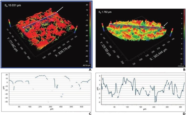
Ultimately, three breast biopsy markers—Cork, Q, and MRI (Flex)—exhibited actionable ultrasound twinkling for ≥2 transducers in cadaveric breast. Additionally, surface roughness was significantly higher for markers with than without actionable twinkling for the C1-6 (median values: 0.97 vs 0.35, p=.02) and 9L (1.75 vs. 0.36; p=.002) transducers.

“The use of twinkling artifact to help detect biopsy markers by ultrasound could impact breast radiologists in performing preoperative localization procedures after NST,” the authors of this AJR article concluded, thus impacting breast surgeons in performing intraoperative localization.

For more information: www.arrs.org


Related Content

News | Magnetic Resonance Imaging (MRI)

Feb. 5, 2026 — Eyas Medical Imaging, Inc. has received U.S. Food and Drug Administration (FDA) 510(k) clearance for its ...

Time February 06, 2026
arrow
News | Ultrasound Women's Health

Feb. 5, 2026 — BrightHeart, a global provider of AI-driven prenatal ultrasound, has announced the availability of its B ...

Time February 05, 2026
arrow
News | Radiation Therapy

Feb. 4, 2026 — On World Cancer Day (02.04.26), the American Society for Radiation Oncology (ASTRO) and the European ...

Time February 04, 2026
arrow
News | Radiology Imaging

Feb. 4, 2026 — The Royal College of Radiologists (RCR) has issued its initial reaction to the British government's ...

Time February 04, 2026
arrow
News | FDA

Feb. 2, 2026 — Imagion Biosystems, Ltd. has submitted an Investigational New Drug (IND) application with the U.S. Food ...

Time February 02, 2026
arrow
News | Breast Imaging | Washington University

Jan. 22, 2026 — In breast cancer, a biopsy is the only diagnostic procedure that can determine if a suspicious lump or ...

Time January 29, 2026
arrow
News | Radiation Oncology

Jan. 27, 2026 — Researchers at the Icahn School of Medicine at Mount Sinai, in collaboration with other leading ...

Time January 29, 2026
arrow
News | Radiology Education

Jan. 22, 2026—The American Roentgen Ray Society (ARRS) will host a live virtual symposium, "Medical Imaging for ...

Time January 28, 2026
arrow
News | Point-of-Care Ultrasound (POCUS)

Jan. 27, 2026 — Siemens Healthineers and World Athletics have joined forces to inform medical teams how point-of-care ...

Time January 27, 2026
arrow
News | Breast Imaging

Jan. 027, 2026 — QT Imaging and Olea Medical have announced plans to collaborate. This collaboration enhances QT Imaging ...

Time January 27, 2026
arrow
Subscribe Now