July 13, 2022 — KA Imaging, a company that develops innovative X-ray imaging solutions, announced the initial results from a study examining the diagnostic value of single-exposure dual-energy subtraction radiography in lung lesion detection.
In the study, a radiologist blinded to standard-of-care CT results was first asked to identify anomalies in the conventional X-ray. The same task was given right after that, with supplemented single-exposure dual-energy images. Quoting directly from the poster, “lesion visibility reportedly increased in 45% of the cases when supplemental dual-energy images were included1.” Findings were validated using CT.
“Lung cancer is the leading cause of cancer death; unfortunately, statistics show that it is rarely detected early,” explains Karim S. Karim, CTO of KA Imaging. “The promising results from this trial show that spectral images can play an important role in earlier detection for better outcomes,” says Amol Karnick, President and CEO.
The study will be presented in an e-poster at this year’s European Congress of Radiology (ECR). In addition to the e-poster at the scientific congress, KA Imaging is part of the technical exhibition at Expo X4, booth 406.
ECR is one of the largest medical meetings in Europe and the second-largest radiological meeting in the world. ECR attendees span all areas of the radiology arena including radiology professionals, radiographers, physicists, industry representatives, and press reporters for both the medical and consumer press.
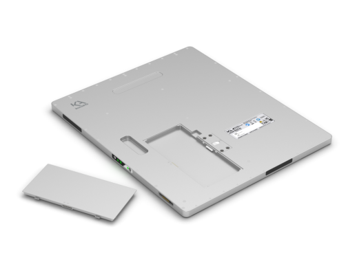
Recently, KA Imaging announced that its patented single-exposure dual-energy technology is now branded SpectralDR. The SpectralDR technology enables dual-energy subtraction, providing bone and tissue differentiation with a single standard X-ray exposure. It acquires three images simultaneously (DR, bone and soft tissue dual-energy X-ray images). The technology mimics the workflow, dose and techniques of state-of-the-art mobile DR X-ray detectors.
For more information: www.kaimaging.com
 November 03, 2026