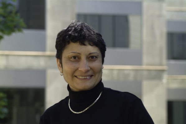
Magda El-Shenawee, University of Arkansas. Image courtesy of University of Arkansas
October 11, 2021 — Electrical engineering professor Magda El-Shenawee’s effort to develop a more accurate and less-invasive method for detecting breast cancer will benefit from a $424,545 grant from the National Institutes of Health.
El-Shenawee works with pulsed, terahertz imaging, a type of electromagnetic radiation technology that produces high-quality images of biological tissue down to roughly 80 micrometers. The method scatters fewer waves than radiography, which enables deeper imaging into the tissue.
Terahertz imaging shows great promise as an alternative method for helping clinicians determine whether all cancerous tissue was removed during and immediately following a lumpectomy.
Standard breast cancer imaging techniques, such as radiography and computed tomography, or CT scan, do not always provide a clear assessment of breast tissue on the margins of a tumor. This is especially important during a lumpectomy, which is the removal of cancerous breast tissue while trying to preserve healthy tissue surrounding it. Without an accurate picture of the margins between the tumor and healthy tissue, surgeons cannot be sure they removed all cancerous tissue.
This problem leads to a high rate — greater than 30 percent — of additional surgery. The need for an immediate imaging technology is especially critical at small hospitals and outpatient clinics that do not have access to an on-site pathology laboratory that could provide immediate results.
“Our pre-clinical models showed strong differentiation between cancerous and fatty tissues,” El-Shenawee said, “but the more clinically relevant differentiation between cancerous and healthy, non-fatty tissue remains challenging. To build upon the successes of our previous work and improve the sensitivity of terahertz imaging to detect cancer at the surgical margins, we have identified areas where we can make significant improvements.”
El-Shenawee’s research team, including Narasimhan Rajaram, associate professor of biomedical engineering, and Jingxian Wu, professor of electrical engineering, in addition to graduate students, will re-design instrumentation to develop more sensitive wave polarization. In this new approach, all four polarizations of waves will be incorporated to increase the spatial and spectral information about different types of tumor tissues.
The researchers will test the system on animal models with breast cancer and try to improve the accuracy of detection algorithms by combining spatial information embedded in the images with spatial statistics.
“We anticipate that the new approach will increase the image contrast between cancerous and healthy adjacent tissues, leading to better differentiation and classification of cancer on the tumor margins,” El-Shenawee said. “The success of this approach should allow us to expand our work and move toward clinical trials.”
For more information: www.uark.edu
 October 19, 2026