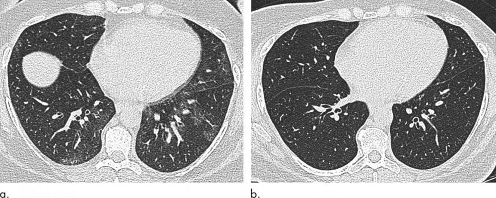
Axial unenhanced inspiratory CT images of the lungs in 51-year-old woman (a) before and (b) 6 months after bariatric surgery with 31-kg weight loss (body mass index decrease, 36.1%). The mosaic attenuation seen before surgery resolved after surgery. Image courtesy of Radiological Society of North America (RSNA)
January 29, 2020 — Bariatric surgery and weight loss appear to reverse some of the negative effects of obesity on the respiratory system, according to a study published in the journal Radiology.
Obesity is a public health epidemic that contributes to a higher risk of hypertension and stroke, diabetes and certain cancers. It also harms the respiratory system, although the scope of these effects is not fully understood.
Known effects of obesity on the respiratory system include increased respiratory work, along with compromised airway resistance and respiratory muscle strength, which may all contribute to restrictive pulmonary function impairment.
As an imaging technology that provides detailed pictures of the lungs and airways, computed tomography (CT) has great potential to improve understanding of obesity’s impact on the respiratory system. Up until now, however, there have been few CT studies evaluating obesity’s effects on the lungs and the trachea, often referred to as the windpipe.
Study lead author Susan J. Copley, M.D., saw CT’s potential firsthand in her practice as a thoracic radiologist at Hammersmith Hospital in London, part of Imperial College Healthcare NHS Trust, where she observed differences on chest CT images obtained in obese individuals.
“This caused me to wonder if these differences were due to obesity and whether they were reversible after weight loss,” she said.
For the study, Copley and her colleagues evaluated changes in the respiratory systems of 51 obese individuals who underwent bariatric surgery, a treatment for obese patients who haven’t responded to other weight loss approaches. The procedure reduces the size of the stomach. All participants lost weight post-surgery with a mean body mass index decrease of 10.5 kg/m2.
The researchers used CT to measure the size and shape of the trachea and assess air trapping, a phenomenon in which excess air remains in the lungs after exhaling, resulting in a reduction in lung function. Air trapping is an indirect sign of obstruction in the small airways of the lung.
When the researchers compared results at baseline and six months after bariatric surgery, they found that surgery and weight loss were associated with morphological, or structural, changes to the lung and trachea.
Post-surgery CT showed reductions in air trapping and a lower incidence of tracheal collapse. Change in the extent of CT air trapping was the strongest predictor of improvement in dyspnea, or shortness of breath.
“For the first time, this study has demonstrated changes in the CT morphology of large and small airways that improve when individuals lose weight,” Copley said. “These features correlate with an improvement in patient symptoms.”
The results suggest that there may be a reversible element of small airway inflammation related to obesity and that reversal of this inflammation correlates with improvement in symptoms. The findings also point to CT as a potential marker of this inflammation.
While more studies are needed to better understand the link between CT features and biomarkers of inflammation, the study underscores CT’s potential in the work-up of patients with obesity.
“CT is a useful morphological marker to demonstrate subtle changes which are not easily assessed by lung function alone,” Copley said.
For more information: www.rsna.org

 July 23, 2026


