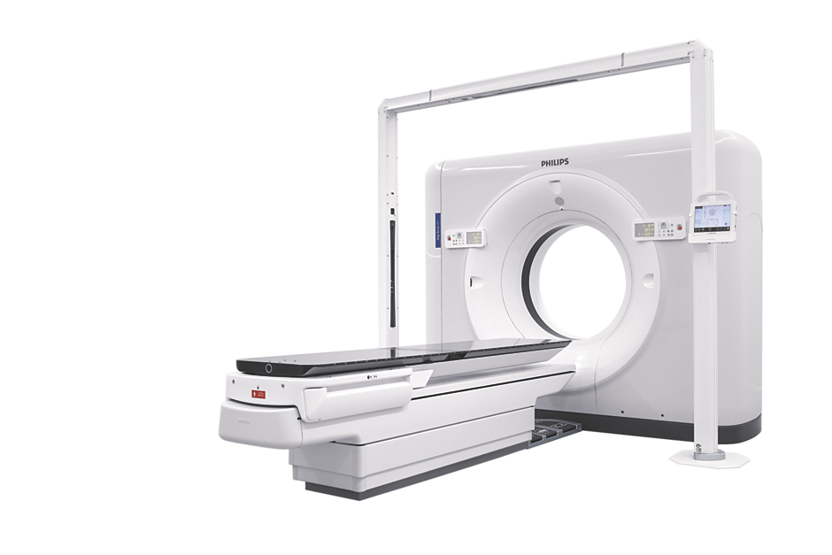
Image courtesy of Philips Healthcare
There were several interesting new trends in cardiovascular computed tomography (CT) imaging at the 2019 Society of Cardiovascular Computed Tomography (SCCT) meeting in July. Key topics at this year’s meeting included integration of artificial intelligence (AI) into CT systems, and the integration of CT calcium scoring into the 2018 American Heart Association (AHA) cholesterol management guidelines. This was according to SCCT President Ron Blankstein, M.D., director of cardiac computed tomography, Brigham and Women’s Hospital, and associate professor of medicine and radiology, Harvard Medical School.
The meeting attendees were about evenly divided between radiologists and cardiologists. Following are four trends that will affect both subspecialties.
1. Artificial Intelligence Enters CT
Vendors are developing new AI algorithms to reconstruct CT images better than conventional iterative or model-based reconstruction methods. Both Canon Medical Systems and GE Healthcare showed examples of their deep-learning CT image reconstruction software, both of which gained U.S. Food and Drug Administration (FDA) clearance in the spring of 2019.
In deep learning, you feed the system the answers (in the case of CT, what ideal clinical images and resolution should look like) and the machine figures out what needs to be done with input image data to make it look like the ideal reference image, explained Jeannie Yu, M.D., FACC, FSCCT, director of cardiovascular imaging, Long Beach VA Health System. In this way, she said, deep learning can be used to help in image resolution recovery that has better noise reduction and resolution than interactive reconstruction images.
Her center is using Canon Medical Systems’ AiCE deep learning reconstruction (DLR) software for the Precision CT system. The technology uses 10 convolutional neuro network (CNN) algorithms with different kernel sizes to reconstruct the images. Yu illustrated that this AI-driven software takes a lot of computing power, with the system operating using 71.2 teraflops. A teraflop is a unit of computing speed equal to one million floating-point operations per second. For comparison, Yu said IBM Watson AI software operates at 80 teraflops, a Playstation 4 game console at 1.8, Xbox One X gaming console at 6 and the iPhone X runs at 5 teraflops.
GE Healthcare also showed examples of its recently cleared Deep Learning Image Reconstruction (DLIR) software on the new Revolution Apex CT system. This next-generation image reconstruction option uses a dedicated deep neural network (DNN) to generate what GE calls TrueFidelity CT Images. Compared to current iterative reconstruction technology, TrueFidelity CT images can elevate every image to offer better image quality, image sharpness and noise texture, according to GE.
2. CT Calcium Scoring in Guidelines
The American Heart Association (AHA) released updated cholesterol management guidelines late last fall that now encourage the use of CT calcium scoring risk assessment. The guidelines state calcium scans can be used to assess a patient’s risk of having a coronary event. This risk assessment also can help patients and their doctors decide if statins should be prescribed. If a patient is on the fence or resistant to taking statins, thought leaders at SCCT say the calcium scan can be used to show the need (or no need) for statins. If a patient has a calcium score of zero, they do not have a risk for coronary events and there is no need for statins.
A low-dose CT calcium scoring scan can be performed to show the amount of calcified atherosclerosis in a patient’s coronary vessels. Software automatically tallies the percentage of plaque in each vessel segment and compiles an Agatston score showing the risk of having a coronary event. Many hospitals now offer calcium scans at a low cost, between $50-100, to screen patients and bring higher-risk patients into that cardiac care network.
3. New CT Scanner Technology
Siemens showed its Go.Top CT scanner. It has a compact CT gantry so it can fit in small rooms and is aimed at cardiology office-based imaging. The system has removable tablets on each side of the scanner which the tech can use to adjust the machine, review scout scans and trigger the scanner. The idea is to improve workflow and allow the tech to remain with the patient at the bedside longer, rather than tucked away in a remote control room using an intercom.
The entire system is built into the gantry, so there is no need for extra equipment in a closet, cabinet or server tower. It comes in a 128-slice configuration with 4 cm of anatomical coverage per rotation. It uses the Stellar detector and tin filtration to eliminate low-energy photons and help lower dose. It can be programmed to aid workflow by automatically removing bone, create curved planar reconstructions, lung computer-aided detection (CAD) and other post-processing features to save time.
4. Advances in Spectral Imaging
Spectral CT has gained a lot of interest in the past few years because of its potential to offer additional information from CT images. This includes the ability to help definitively make a diagnosis in cases of small pulmonary embolisms, determining if shadowing inside a stent is artifact or restenosis, better delineation of aortic stent graft endoleaks, and to better visualize ischemia or infarcts using enhanced iodine mapping techniques based on different X-ray energies, explained outgoing SCCT President Suhny Abbara, M.D., FSCCT, in sessions. He is chief of cardiothoracic imaging and chair of the CT operations committee, University of Texas Southwestern Medical Center, Dallas, and is heavily involved in spectral imaging research.
Siemens, GE, Philips and Canon now all offer spectral imaging capabilities on some of their newest scanners.

 July 20, 2026


