June 8, 2016 — The presence of high saturated fatty acids in breast tissue may be a useful indicator of cancer in postmenopausal women, according to a new study by researchers at NYU Langone Medical Center. The study was published online in Radiology, a journal of the Radiological Society of North America.
Specifically, the researchers used a new technique developed in NYU Langone’s Department of Radiology that helps identify the relationship between fatty acids and breast cancer. These findings, they say, may one day lead to greater understanding of the underlying mechanisms behind breast cancer development and the role of fat as a factor in breast cancer diagnosis and progression.
“Our study offers the first evidence — seen in breast tissue — that high saturated fatty acids in the breast adipose tissue is associated with presence of breast cancer in postmenopausal women,” said senior author and investigator Sungheon G. Kim, Ph.D., associate professor in the Department of Radiology at NYU Langone and a researcher at the Center for Advanced Imaging, Innovation and Research.
The relationship between body mass index (BMI), fat and cancer development has previously been studied, with postmenopausal women found to be at increased risk for breast cancer as their BMI increases. However, this study suggests the composition of the fat itself may play a role as well.
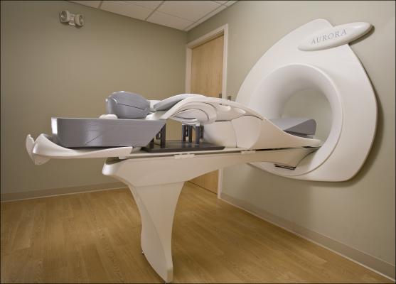
In order to measure fat composition, the NYU Langone radiology research team developed a new approach to magnetic resonance (MR) spectroscopic imaging — a type of MRI that provides information on the chemical composition of the tissue. Kim and colleagues devised a novel method called gradient-echo spectroscopic imaging that provides information on various types of fatty acids based on a series of three-dimensional MRI images acquired for five minutes.
“There is a clear need for methods that can accurately measure fat composition of the breast tissue within a short scan time, and our study takes a first step towards meeting this critical gap,” said Kim.
In this study, researchers analyzed imaging sequences from a total of 89 women who were, on average, approximately 48 years old. Fifty-eight women were pre-menopausal and 31 women were post-menopausal. Each patient’s height, weight and BMI were recorded.
All women received an additional five-minute scan of three-dimensional multiple gradient echo sequences at the end of their diagnostic MRI exams. Forty-nine patients had benign breast tissue, 12 had ductal carcinoma and 28 had invasive ductal carcinoma.
Compared to the benign breast tissue of postmenopausal women, results showed that the breast tissue in postmenopausal women with invasive ductal carcinoma was comprised of a higher percentage of saturated fatty acids and a lower percentage of monounsaturated fatty acids. These findings suggest high-saturated fatty acids and low monounsaturated fatty acids may be associated with invasive cancer, according to the study’s authors.
Of the women with benign lesions, postmenopausal women exhibited higher polyunsaturated fatty acids and lower saturated fatty acids than the premenopausal women.
No significant correlation was found between BMI and fatty acids in breast tissue, suggesting that data regarding the composition of fats could be more indicative of breast cancer.
Because the population screened in this study was considered a high-risk group, and they were scheduled for high-risk screening follow-up, or suspicion of cancer, more research is needed to determine the role of fat composition in low-risk postmenopausal women, the authors say. They also point out that further research also is needed to determine how these fats, which are created in the body and are not correlated with dietary intake, may influence cancer development.
“Measuring breast fat composition only takes an extra five minutes, making this practical, new technique something that could easily be implemented in a clinical setting,” said study co-author Linda Moy, M.D., an associate professor in the Department of Radiology at NYU Langone and a member of its Laura and Isaac Perlmutter Cancer Center. “With further research, we could potentially use these findings to change how we look at breast cancer imaging.”
Funding support for the study, which took fourteen months to complete, was provided by NIH grant R01-CA160620.
Besides Kim and Moy, other NYU Langone researchers involved in this research study at the time were co-investigators Melanie Freed, Ph.D.; Pippa Storey, Ph.D.; Alana Amarosa Lewin, M.D.; Jim Babb, Ph.D.; and Melanie Moccaldi, RT.
For more information: www.pubs.rsna.org/journal/radiology

 July 07, 2026


