What’s healthy? What’s not? These questions should be easy to answer in our world of high-tech, high-resolution scanners. But population-based studies demonstrate they are anything but.
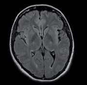
A magnetic resonance (MR) brain scan of a normal volunteer in one such study showed multiple microbleeds and white matter lesions.
“If this were a patient, I would say it was a stroke,” said Norbert Hosten, president of the German Radiological Society, speaking to journalists at a press conference at the European Congress of Radiology (ECR). But the volunteer showed no clinical signs of a stroke — or any other disease, said Hosten, who cited a multitude of potential gains that might come from population-based imaging.
“With the imaging of normal volunteers, we might quantify the normal amount of fat in the liver; find out what a normal joint looks like,” he said. “Studies of normals can revolutionize how we look at images.”
Hosten related the case of a normal volunteer whose MR of the spine showed a slipped disk. “If we had a patient present at the clinic with the same images, we might have operated on him,” he said.
The need for population-based research is compelling, Hosten said. Radiologists need to know the range of what is normal. MRI, for example, is being widely applied in the diagnosis of breast cancer. “Yet very few normal women have been studied, so we don’t know what a normal breast looks like,” he said.
Studies of presumably healthy people can be done ethically with imaging modalities that pose no risk from ionizing radiation. Europe has many such studies underway. On the north shore of the Baltic Sea, bordering Poland and Germany, radiologists are performing free whole-body MR scans of healthy subjects. Tissues and structures throughout the body are being examined and documented.
The Dutch have performed several population-based studies over the past two decades. One looked at more than 10,000 normal volunteers near Rotterdam with the aim of determining whether MR might pick up early signs of neurodegenerative diseases.
Cardiology and oncology may gain remarkably as this kind of research might illuminate early signs of cardiac pathology and tumor development. The same may be true for virtually any human condition, Hosten said, from liver cirrhosis to osteoporosis.
While the use of nonionizing radiation eliminates risk from radiation that might otherwise be needed for imaging, population-based research is not without controversy. The examination of presumably healthy individuals begs the question: What responsibility does the radiologist have when uncovering apparently pathologic conditions in seemingly healthy individuals?
In a traditional epidemiological study, healthcare providers do not intervene, Hosten said. But with modern imaging technology, some signs of disease may demand action. An example is the discovery of a kidney tumor.
“We know what a kidney tumor looks like, so if you do an MR scan and spot such a tumor, I think you are obliged to intervene,” Hosten said.
But where do researchers draw the line? No action was taken, for example, in the patient who showed microbleeds and white matter lesions. What is so obvious that the radiologist has a duty to step in?
“That is the big question,” Hosten said, “when do we and when do we not intervene?”
Greg Freiherr has reported on developments in radiology since 1983. He runs the consulting service, The Freiherr Group. Read more of his views on his blog at www.itnonline.com.

 July 10, 2026


