October 14, 2011 — A study published in the September/October issue of Radiologic Technology shows increasing the source-to-image distance for direct digital radiography (DR) pelvic examinations reduces radiation dose while maintaining image quality. RT is a journal of the American Society of Radiologic Technologists (ASRT).
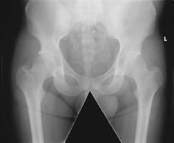
Researchers from the United Kingdom investigated distance, dose and image quality for DR pelvic exams, the second most common Bucky procedure in the country.
To perform the study, the researchers positioned an anthropomorphic pelvic phantom for a standard anteroposterior examination. Using a single DR unit with a flat panel detector, they set an initial source-to-image distance of 100 cm and used an antiscatter focused radiation grid. The x-ray beam was collimated for a standard AP pelvic examination and was kept constant throughout the experiment, while source-to-image distance varied from 80-147 cm.
Two exposures were taken in 10-cm intervals to determine image quality, and the experiment was repeated without the grid. A total of 80 images were then sent to a reporting-grade picture archiving and communication workstation for image quality analysis. Researchers calculated the entrance surface dose and effective dose based on distances of 60 cm, 80 cm, 100 cm, 120 cm, 140 cm and 147 cm.
The research results indicate that by increasing the source-to-image distance, both the entrance surface dose and effective dose decreased when using the antiscatter radiation grid; they decreased further when the grid was removed. At 147 cm, the decrease in entrance surface dose and effective dose when using a grid equated to a 25 percent reduction in radiation dose compared with standard parameters.
“The optimization of radiation dose for both computed radiography and digital radiography examination is an essential part of radiographic practice,” said Andrew England, BSc(Hons), PgCert, MSc, FHEA, a study author and lecturer for the directorate of medical imaging and radiotherapy at the University of Liverpool. “Our lab-based phantom experiments have indicated that dose reduction is possible without loss of any image quality.”
In addition, the findings suggest that as source-to-image distance increases, there also may be a minor increase in image quality, although it was determined to be statistically insignificant.
Based on the study’s outcome, researchers recommended a clinical study involving patients is necessary to confirm the dose reduction potential of increasing source-to-image distances. “It is essential that any improvements to radiographic technique are carefully evaluated in a clinical setting as well as in the lab before we can promote the widespread adaption of any new techniques,” England said.
For more information: www.asrt.org

 June 23, 2026


