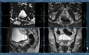
Multiplanar T2-weighted images and ADC map.
Prostate disease is primarily evaluated by using digital rectal examination (DRE) in combination with other clinical data including prostate-specific antigen (PSA) tests and pathologic information. These commonly used diagnostic tests have been shown to result in both high false positive and high false negative results. Additionally, the use of transrectal ultrasound-guided (TRUS) biopsy techniques for cancer detection can miss 20 percent or more of prostate cancers.
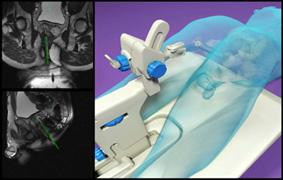
The use of DynaCAD for Prostate for image analysis and DynaTRIM (Trans-Rectal Interventional MRI) is a new option for evaluation of prostate disease, providing radiologists and urologists with another tool to diagnose their patients.
Clinical Case Study With Elevated PSA
A 67-year-old man presented with a PSA level of 5.4 ng/ml and no palpable abnormality on DRE. The first of two TRUS biopsies was performed with eight core samples. No malignancy was found. Due to elevated PSA, follow up was recommended.
Six months later, the second TRUS biopsy was performed with 12 core samples. Again, no malignancy was found and follow up was once again recommended. DRE was also performed with no palpable abnormalities. Since his PSA levels remained elevated, the patient was referred for a diagnostic MRI study. The examination was performed on a 3.0T MRI scanner with the body array coil in a dual-coil setup. Multiparametric imaging sequences were acquired including high-resolution, multiplanar T2-weighted anatomical images.
Diffusion-weighted images with b-values of 50, 500 and 800 were acquired and an ADC map was generated. The study was then imported to DynaCAD for Prostate for analysis and interpretation.
Image Results: A suspicious area was identified on axial T2. Based on these findings, MR-guided biopsy was recommended. MR-guided prostate biopsy was performed with DynaCAD for Prostate and DynaTRIM.
Diagnosis: Confirmed prostate pancer - Gleason 7 (75 percent in one core).
Summary: This patient, presenting with an elevated PSA, was able to benefit from this new imaging and interventional option where multiple TRUS-guided biopsies were performed with negative results. The clinician utilized DynaCAD for Prostate for the image analysis, which drew attention to suspicious areas and DynaTRIM for the targeted biopsy which resulted in a confirmed diagnosis.
This case study was supplied by Invivo Corp.
For more information: www.invivo.com, www.admetech.org and http://urology.jhu.edu/newsletter/prostate_cancer58.php

 October 07, 2026