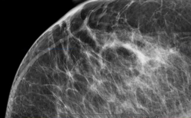
April 15, 2014 — VuComp Inc. announced it has released an updated version of its M-Vu Breast Density technology. The new M-Vu Breast Density 2.0 adds a critical dimension to the analysis of dense breast tissue. The VuComp density category, analogous to the BI-RADS breast density composition category, is now correlated to not only the amount, but also the distribution – the actual dispersion – of fibroglandular tissue.
M-Vu Breast Density automatically and rapidly assesses breast density and provides consistent and accurate measurements. The tool evaluates mammograms by analyzing the structure, texture and dispersion of the tissue, rather than simply estimating total fibroglandular volume. To ensure product effectiveness, the findings of a panel of 13 expert radiologists were used to calibrate the VuComp density categories to the American College of Radiology’s (ACR) Breast Imaging Reporting and Data System (BI-RADS) standard. The M-Vu algorithms quantify areas with a dense appearance that could hide cancer, and convert them to categories corresponding to the recently updated standard. This exclusive approach using appearance-based texture and dispersion analysis provides useful adjunctive information. M-Vu Breast Density is the only commercially available, U.S. Food and Drug Administration (FDA) -cleared system that employs this scientific methodology.
Clinical studies have shown that dense breasts can hide a cancerous lesion by reducing the ability to visualize fine structures and details that could indicate a malignant abnormality, making breast cancer detection in a mammogram more difficult. Patients with dense breast tissue may be recommended for additional screening exams. Legislation has passed in 16 states requiring physicians to notify patients if their breast density is in one of the two higher categories.
“VuComp understands that breast density measurement is deeper than just an analysis of volume,” said Jim Pike, president of VuComp. “We are dedicated to continual improvements in the science of automated density and believe that radiologists will recognize the clinical utility of this tool to be even more useful to their practice than before. We are very pleased that all existing M-Vu Breast Density users will have this important product enhancement available immediately.”
For more information: www.vucomp.com

 July 07, 2026


