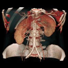
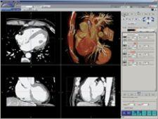
Advanced cutplane feature is able to expose LAD from AquariusNet thin-client application.
In the early 1990s, while my colleagues and I at Stanford University foresaw the power of volumetric data as a logical extension of spiral CT evolution, we did not foresee the necessity of these tools to efficiently navigate and interpret CT scans composed of thousands of transverse reconstructions.
Over the years, however, real-time volumetric CT interactivity became a logical extension of the evolution of CT acquisition. CT scanning had evolved into a modality that was no longer limited to transverse planar reconstructions, but to a volumetric technique best examined with the full spectrum of planar and volumetric display techniques available.
Real-time interactivity with 2,000 to 4,000 image CT datasets using volume rendering requires dedicated hardware platforms with specialized graphics processing units, but allowing for widespread availability to these workstations can be costly. A system capable of distributing computing and graphics power emerged as the logical solution for efficient and cost-effective delivery of distributed 3-D visualization capabilities.
In 2002, Stanford University acquired such a system, the AquariusNET server-client visualization system from TeraRecon.
By setting the single server in a convenient location within the medical center, we were instantly able to access it from any point within our medical center’s network and interact with patient data using high-quality, real-time volume rendering from any location accessible to a network connection. Moreover, the sophisticated visualization and interactions were possible with the simplest personal computers — laptops or desktops. Within days, 3-D visualization was available everywhere throughout the medical center, not just in two or three reading rooms.
I believe the introduction of this system has ushered in the widespread use of 3-D visualization for primary data interaction by radiologists and clinicians within our medical center. Prior to its introduction, the 3-D workstation was viewed as a tool for the dedicated 3-D technologist’s use. It has now become our viewbox. I am impressed daily by the added information that we extract from real-time 3-D interactivity when compared to primary transverse section review.
The greatest value of this technology, however, emerges when clinical colleagues arrive to discuss the complexities of their patients’ problems. We are are able to distill 4,000 CT images into a simple interactive exploration that fully communicates the information needed for those clinicians to deliver their best care to their patients.
Our clinical conferences have evolved into an entirely new level of discussion and interaction. An LCD projector and a laptop running the AquariusNET Client software enables us to access any recently studied patient to fully explore the subtleties of their disease distribution, achieving the synergy that should result when physicians with varied specialties come together to optimize a patient’s care.
Note: This case study is an excerpt from Dr. Rubin’s original
article, which can viewed in its entirety at www.terarecon.com/downloads/news/article_rubin.pdf.

 April 18, 2025