Dec. 12, 2024 — Lunit, a provider of AI-powered solutions for cancer diagnostics and therapeutics, recently announced the publication of a new study in npj Precision Oncology detailing the development of its Universal Immunohistochemistry (uIHC) AI model.
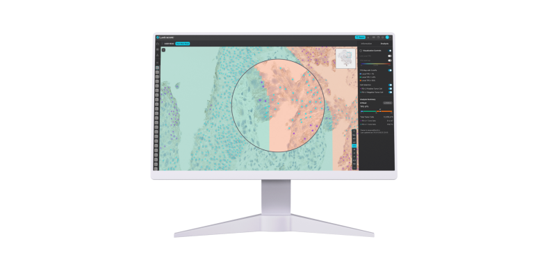
The study demonstrates how the model excels at analyzing diverse cancer types and IHC stains, including datasets it had never encountered before, due to a novel training approach. Now commercialized as Lunit SCOPE uIHC, the model enables advanced biomarker formation from even singleplex IHC, with subcellular stain localization, continuous intensity scoring, and cell type identification.
Addressing the Challenges
Immunohistochemistry (IHC) is an essential tool in oncology, enabling pathologists to detect and quantify protein expression which in turn guides decisions for systemic therapy. However, while several AI algorithms exist to assist in scoring IHC images and improving accuracy, current AI models face two major limitations:
1. Data Dependency: Current AI-IHC models require large numbers of immunostain-specific images for training, which are difficult to obtain, particularly for novel immunostain-target pairs.
2. Lack of Generalization: Current AI-IHC models struggle to analyze datasets that differ from their training set either in immunostain or cancer types, limiting their ability to be effective in diverse indications.
These challenges underscore the need for scalable solutions capable of accurate analysis across a wide range of cancer types and immunostains.
uIHC Model
Lunit’s study compared eight deep learning models, including four single-cohort (trained using data from a single stain or cancer type) and four multi-cohort (trained on combined datasets spanning multiple stains and cancer types) approaches, to evaluate their performance on both familiar and unseen datasets. The results validated the uIHC model’s ability to generalize across diverse datasets with high accuracy.
Key results include:
- High Concordance on Known Datasets: The uIHC model achieved a Cohen’s kappa score of 0.792, surpassing the best single-cohort model, which scored 0.744 when analyzing known cancer types and immunostains.
- Superior Generalization to Unseen Data: On novel datasets involving previously unseen cancer types and immunostains, the uIHC model achieved a Cohen’s kappa score of 0.610, representing a relative improvement of 10.2% over the single-cohort model average of 0.508.
- Enhanced Tumor Proportion Score (TPS) Accuracy: Across multi-stain test sets, the uIHC model achieved an AUC of 0.921 for TPS evaluations and a TPS accuracy of 75.7%, demonstrating its reliability in quantifying IHC images.
These findings highlight the model’s robust performance across a wide variety of cancer types and immunostains, including those it had not been trained on.
The uIHC model’s ability to generalize across diverse IHC images marks a transformative step in digital pathology. By reducing the dependency on large stain-specific datasets, it enables scalable and efficient biomarker analysis for clinical diagnostics and drug development. This capability is particularly valuable for evaluating new biomarkers associated with novel therapies, addressing a critical bottleneck in precision oncology.
“Our Universal Immunohistochemistry AI model solves a practical bottleneck in development settings—handling unseen cancer types and stains without requiring additional data annotation,” said Brandon Suh, CEO of Lunit. “By proving the effectiveness of a multi-cohort training approach, this study shows how AI can be adapted to real-world complexities, delivering both precision and scalability. With the launch of Lunit SCOPE uIHC, we’re enabling researchers and clinicians to focus on what truly matters: advancing patient care and accelerating therapeutic innovation.”
More information is available at lunit.io.

 July 09, 2026


