March 24, 2021 — RSIP Vision, a leading innovator in medical imaging through advanced artificial intelligence (AI) and computer vision solutions, announced an advanced joint segmentation tool for detailed, non-invasive planning of revision arthroplasty and other orthopedic procedures for patients with pre-existing metal implants. This powerful AI-based software module enables quick and accurate segmentation of different joints from computed tomography (CT) scans of hips, knees, shoulder and spines. It provides precise measurements of the geometry of joints, including complicated cases of joints with existing metallic orthopedic implants. RSIP Vision’s deep learning algorithms provide a solution for the artifacts associated with metals in CT images, which normally cause severe degradation of medical imaging. This vendor-neutral technology will be available to third-party CT manufacturers and medical device vendors, allowing them an improved way to plan and execute both manual and robot-assisted revision arthroplasty procedures.
“One of the most common problems in bone segmentation imaging occurs when a CT scan includes the presence of metals in the bones, either due to previous orthopedic procedures (such as hip replacements) or surgical corrections after a traumatic injury,” said Ron Soferman, CEO of RSIP Vision. “In these cases, the CT images become problematic because of nonstandard absorption values caused by crosstalk between the absorbing pixels and additional artifacts — which result in a challenging image. Moreover, standard segmentation tools can lead to inaccurate outcomes that limit the orthopedic surgeon’s ability to properly plan for the surgery.”
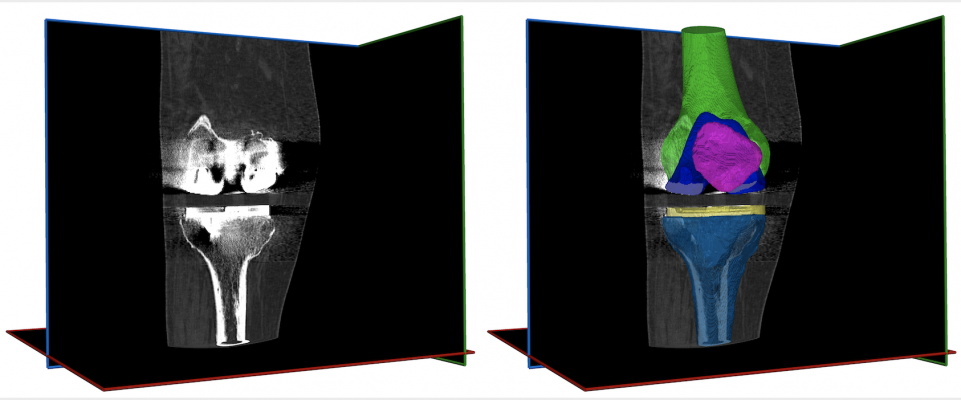
RSIP Vision’s innovative module uses a state-of-the-art deep neural network approach, designed and trained in advance to deal with the presence of metals in CT scans. This module features accurate discernment of the location of metals in the bones, resulting in uniform, accurate and robust segmentation of all the different elements of both bones and implants, as well as exact delineation of metal fragments from the bones. The technology allows the surgeon to plan in an optimal manner. This leads to better preparation and setting of expectations with the patient. In addition, preparing the required implants rather than all the various options makes it more financially beneficial.
This technology may be used in a wide variety of orthopedic use cases, including periprosthetic fractures around implants or fusions, local recurrence of tumors in the presence of implants and repeat femoral neck fracture after fusion.
“This module is a vital and important contribution to the treatment of patients undergoing orthopedic procedures since many patients undergo additional or follow-up procedures throughout their lifetime,” said Soferman.
“The advantage of bones and implant segmentation in CT scans is the surgeon's ability to accurately evaluate the bone structure, including significant bone loss in order to optimally prepare for revision surgery,” said Shai Factor, M.D., Orthopedic Surgery Resident, Department of Orthopedic Surgery Tel Aviv Medical Center, Israel. “Assessment and preparation for revision surgery when the condition of the bone surrounding the implant is known in advance enables us to make pre-surgical decisions including which new implant to use and the degree of incision required. This will allow for ordering the desired implant in advance, as well as preparing the patient for the expected outcome and the accompanying rehabilitation process.”
For more information: www.rsipvision.com

 June 25, 2026


