Image courtesy of Planmeca
March 4, 2016 — The first facial tissue transplant procedure in the history of the Nordic countries was performed earlier this year in the Hospital District of Helsinki and Uusimaa (HUS) in Finland. Planmeca contributed to the demanding and rare operation, which was the 35th of its kind in the world to date.
The facial tissue transplant surgery itself took 21 hours and was carried out by a group of 11 surgeons, as well as 20 nurses and other experts. The operation consisted of transplanting the patient’s upper and lower jaw, lips and nose, as well as segments of their skin, midfacial and tongue muscles, and the nerves of these muscles. The head the surgical team, Patrik Lassus, M.D., emphasized that the objective of the operation was to transplant facial functions, not external features.
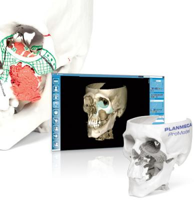
The Planmeca ProModel service was part of the demanding procedure. It is a service for designing and creating patient-specific implants, surgical guides and skull models from cone beam computed tomography (CBCT)/CT images. 3-D technology decreases surgical time and produces significantly more precise results when compared to traditional methods. This makes operations increasingly safer for patients.
The facial tissue transplant procedure was planned pre-operatively utilizing 3-D technology. The planning consisted of modeling donor tissues and determining how they match the recipient. Surgeons Lassus and Jyrki Törnwall, M.D., designed the 3-D-printed surgical guides together with Planmeca’s CAD/CAM designer.
Planmeca’s software substantially decreased the operating time – saving hours compared to similar procedures previously carried out elsewhere in the world. Conserving time is one of the key aspects of surgery, as longer operations increase the risk of complications. In transplant cases, it is also of paramount importance to accelerate the restoration of blood flow.
“Based on literature, we know that it can take 3 to 4 hours to trim bones. In this particular operation, it took Patrik [Lassus] and myself under 10 minutes to place the transplant. This led to a drastic reduction in the duration of the surgery, while also significantly improving the accuracy of bone placement,” described Törnwall in the press conference on the operation.
Planmeca participated in planning the facial tissue transplant right from the start, led by CAD/CAM Design Manager Jani Horelli.
At Planmeca, planning the operation began around three years ago. Careful steps were taken in preparing for the upcoming procedure.
“Planmeca’s part consisted of two phases. First, we designed the surgical guides together with Dr. Lassus and Dr. Törnwall, as well as determined the kinds of segments that would be surgically removed from the recipient and transplanted from the donor. At this point, we were anticipating a scenario, which would become concrete once a donor was found,” recounted Horelli. “The second phase began immediately once we received word of a suitable donor. An X-ray image of the donor was taken at the hospital and the imaging data was utilized in 3-D designing. We also simulated the operation together with the surgeons. Following this, the components were designed and manufactured at Planmeca headquarters and transported to the hospital, where they were taken directly to the operating room.”
“Planmeca’s role has been essential to our work for years – we have been able to utilize computer simulations to create saw guides, which allow us to saw at a specific orientation and to an exact depth, as well as remove facial structures, which we know match the donor, at a precise angle,” Törnwall noted.
For more information: www.planmeca.com

 April 18, 2025