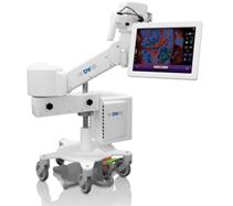
Image courtesy of DySIS Medical
January 19, 2015 — A new computerized diagnostic tool designed to assist in the early detection of cancerous and pre-cancerous cervical lesions, called DySIS (Dynamic Spectral Imaging System) Advanced Cervical Imaging System, is making its U.S. debut after undergoing extensive testing and review in Europe.
Endorsed by the U.K. National Institute of Health and Care Excellence, the new U.S. Food and Drug Administration (FDA)-cleared medical device is being praised by physicians for its ability to help identify abnormalities on the cervix, which can sometimes escape detection during a standard colposcopy.
Colposcopy is a diagnostic exam, usually following an abnormal Pap smear, in which a doctor applies a special solution to the cervix and peers into a binocular viewer called a colposcope to look for areas that turn white, indicating abnormal cells and that a cervical biopsy may be needed.
"The DySIS system is a next generation colposcope, offering important advancements that improve the examination procedure for both doctors and patients," said Christopher G. Olson, M.D., of Women's Center for Health in Naperville, one of the first OB/GYN practices in the United States to offer the new computer-assisted screening device.
The DySIS Advanced Cervical Imaging System is used by the examining physician as follows:
- A high-resolution digital image of the cervix is displayed on the DySIS touchscreen to allow normal assessment of the cervical area. Image color, brightness, contrast and magnification of the displayed image can be adjusted.
- Acetic acid, which turns abnormal cervical cells white, is applied to the surface of the cervix by the physician via a built-in syringe with a diffuser for instant homogenized coverage over the cervix.
- The operator uses the touchscreen interface to conduct a standard colposcopic examination while DySIS measures the acetowhitening effect. Meanwhile, DySIS uses Dynamic Spectral Imaging to record the entire acetowhitening process.
- The system then produces a DySISmap Advanced Cervical Scan, which is like a weather map showing the precise areas of the cervix where acetowhitening is most extreme and most likely to contain abnormal cells.
- The physician refers to the DySISmap and marks biopsy points that may be needed based on the information displayed as well as other examination information.
- The scanned digital image of the cervix and DySISmap showing acetowhitening and biopsy points are then saved along with patient notes entered via the touchscreen.
Olson says that a traditional colposcopy can often be very frightening for a patient because during the examination they have no idea what the doctor is looking at or thinking.
"Thirty seconds of silence can seem like five minutes, and a patient's imagination can begin to race as they assume the doctor has discovered the worst," Olson said.
With the DySIS system, Olson says a patient is able to view what the doctor is viewing in real-time and discuss the meaning of the colorized zones and markings on the DySISmap, enabling the patient to be involved and more at ease during the examination procedure.
"I was skeptical that a computer-generated image could pick up cervical abnormalities better than the naked eye, but I was wrong," Olson said. "On a colposcopic image that appeared normal to me, the DySISmap highlighted two areas of abnormalities revealing cervical dysplasia on biopsy. With the new recommendations to decrease Pap smear frequency, it is even more important that colposcopies miss nothing."
Olson says examinations performed using the DySIS Advanced Cervical Imaging System do not result in any additional out-of-pocket expenses for the patient.
For more information: www.dysismedical.com

 July 02, 2026


