September 15, 2015 — Imaging patients soon after a traumatic brain injury (TBI) can lead to more accurate detection of cerebral microhemorrhages, or microbleeding on the brain, according to a study of military service members. Results were published online in the journal Radiology.
Cerebral microhemorrhages occur as a direct result of TBI and can lead to severe secondary injuries such as brain swelling or stroke. The ability to monitor the evolution of microhemorrhages could provide important information regarding disease progression or recovery.
According to the Centers for Disease Control and Prevention’s (CDC) National Center for Injury and Prevention Control, about 1.7 million people in the United States sustain TBI each year. Furthermore, the Institute of Medicine reports that 20 to 23 percent of military service members deployed to Afghanistan and Iraq have sustained TBI while serving.
“TBI is a large problem for our military service members and their families,” said Gerard Riedy, M.D., Ph.D., chief of neuroimaging at the National Intrepid Center of Excellence at the Walter Reed National Military Medical Center in Bethesda, Maryland. “We found that many of those who have served and suffered this type of injury were not imaged until many, many months after injury occurred, thus resulting in lower rates of cerebral microhemorrhage detection which delays treatment.”
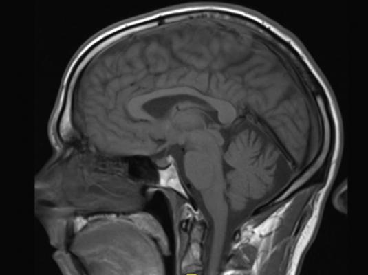
For the study, Riedy and colleagues used susceptibility-weighted imaging—a magnetic resonance imaging (MRI) technique that provides improved visibility of blood and is highly sensitive to hemorrhage—to evaluate 603 military service members with TBI. The median time from point of injury to imaging was 856 days. Of the 603 military service members who participated in the study, 7 percent were found to have at least one occurrence of cerebral microhemorrhage.
The patients were divided into four groups based on time since the injury occurred, ranging from less than three months to over a year. The results found that those who were imaged more than a year after the injury had a much lower occurrence of cerebral microhemorrhages than those who were scanned 12 months or fewer after TBI.
Cerebral microhemorrhage was identified in 24 percent of military personnel who were imaged within three months post-injury, compared to 5.2 percent of the patients who were imaged over a year later. The researchers attribute this to changes in iron deposits in the brain as time goes on, making it more difficult to detect microbleeding.
“Early characterization of cerebral microhemorrhages may help to explain clinical symptoms of acute TBI and identify the severity of brain damage,” Riedy said. “We believe that having access to MRI in the field would facilitate early detection of TBI, thus providing timely treatment.”
The study also supports previous claims that using susceptibility-weighted imaging to evaluate brain injury patients may be more effective than conventional MRI. In this study’s capacity, using susceptibility-weighted imaging resulted in detecting significantly more microhemorrhages due to a higher spatial resolution and signal, with 77 percent of cerebral microhemorrhages appearing more evident through susceptibility-weighted imaging when compared to conventional MRI.
For more information: www.nicoe.capmed.mil

 July 10, 2026


