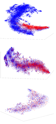
The test images are divided into three subsets. Images with: 11 a) low uncertainty 11 b) medium uncertainty and 11 c) high uncertainty. A dimensionality reduction of the images reveals that the images with low uncertainty (11 a) show clear distinction between the benign and malignant images. These are the images with low uncertainty are easily separable in low dimensions and the machine learning model is confident in classifying these images. Whereas the images with high uncertainty are randomly distributed in three dimensions (11 c). For medium uncertainty images, the images are clustered without a clear distinction of classes. Thus, explaining the uncertainty quantified by the machine learning model.
(Courtesy Ponkrshnan Thiagarajan/Michigan Tech)
December 8, 2021 — A Michigan Tech-developed machine learning model uses probability to more accurately classify breast cancer shown in histopathology images and evaluate the uncertainty of its predictions.
Breast cancer is the most common cancer with the highest mortality rate. Swift detection and diagnosis diminish the impact of the disease. However, classifying breast cancer using histopathology images—tissues and cells examined under a microscope—is a challenging task because of bias in the data and the unavailability of annotated data in large quantities. Automatic detection of breast cancer using convolutional neural network (CNN), a machine learning technique, has shown promise; however, it is associated with a high risk of false positives and false negatives.
Without any measure of confidence, such false predictions of CNN could lead to catastrophic outcomes. A new machine learning model developed by Michigan Technological University researchers, however, can evaluate the uncertainty in its predictions as it classifies benign and malignant tumors, helping reduce this risk.
In a paper recently published in the journal IEEE Transactions on Medical Imaging, mechanical engineering graduate students Ponkrshnan Thiagarajan and Pushkar Kharinar and Susanta Ghosh, assistant professor of mechanical engineering and machine learning expert, outline their novel probabilistic machine learning model, which outperforms similar models.
“Any machine learning algorithm that has been developed so far will have some uncertainty in its prediction,” Thiagarajan said. “There is little way to quantify those uncertainties. Even if an algorithm tells us a person has cancer, we do not know the level of confidence in that prediction.”
From Experience Comes Confidence
In the medical context, not knowing how confident an algorithm is has made it difficult to rely on computer-generated predictions. The present model is an extension of the Bayesian neural network—a machine learning model that can evaluate an image and produce an output. The parameters for this model are treated as random variables that facilitate uncertainty quantification.
The Michigan Tech model differentiates between negative and positive classes by analyzing the images, which at their most basic level are collections of pixels. In addition to this classification, the model can measure the uncertainty in its predictions.
In a medical laboratory, such a model promises time savings by classifying images faster than a lab tech. And, because the model can evaluate its own level of certainty, it can refer the images to a human expert when it is less confident.
But why is a mechanical engineer creating algorithms for the medical community? Thiagarajan’s idea kindled when he started using machine learning to reduce the computational time needed for mechanical engineering problems. Whether a computation evaluates the deformation of building materials or determines whether someone has breast cancer, it’s important to know the uncertainty of that computation—the key ideas remain the same.
“Breast cancer is one of the cancers that has the highest mortality and highest incidence,” Thiagarajan said. “We believe that this is an exciting problem wherein better algorithms can make an impact on people’s lives directly.”
Next Steps
Now that the study has been published, the researchers will extend the model for multiclass classification of breast cancer. They will aim to detect cancer subtypes in addition to classifying benign and malignant tissues. And the model, though developed using breast cancer histopathology images, can also be extended for other medical diagnoses.
“Despite the promise of machine learning-based classification models, their predictions suffer from uncertainties due to the inherent randomness and the bias in the data and the scarcity of large datasets,” Ghosh said. “Our work attempts to address these issues and quantifies, uses and explains the uncertainty.”
Ultimately, Thiagarajan, Khairnar and Ghosh’s model itself—which can evaluate whether images have high or low measures uncertainty and identify when images need the eyes of a medical expert—represents the next steps in the endeavor of machine learning.
For more information: www.mtu.edu


 February 06, 2026


