News | Cardiovascular Ultrasound | July 02, 2019

Konica will incorporate DiA’s LVivo Toolbox for cardiac ultrasound analysis into Exa Cardio PACS

Konica Minolta Healthcare Partners With DiA Imaging Analysis for AI-based Cardiac Ultrasound Analysis

July 2, 2019 — DiA Imaging Analysis has partnered with Konica Minolta Healthcare Americas Inc. to expand analysis capabilities of Konica Minolta's Exa Cardio PACS platform (cardiovascular information system) with DiA's LVivo Toolbox for cardiac analysis.

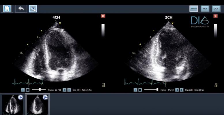
The LVivo Cardiac Toolbox, an artificial intelligence (AI)-powered solution, is designed to analyze cardiac ultrasound images automatically and objectively, to reduce the subjectivity of manual or visual analysis methods used today.

DiA's LVivo Cardiac Toolbox uses novel pattern recognition, deep learning and machine learning algorithms that automatically imitate how the human eye detects borders and motion. DiA's automated solution generates fast and accurate image analysis to support the clinician's decision-making process.

LVivo Cardiac Toolbox is vendor-neutral, supporting DICOM clips of various ultrasound systems. Konica Minolta will offer the LVivo Toolbox as a part of Exa's diagnostic-quality zero-footprint, server side rendering universal viewer for DICOM and non-DICOM images. The integration has been designed according to Exa's user interface to assure the most efficient workflow and accessibility to all Exa Cardio PACS (picture archiving and communication system) users.

Konica Minolta's Exa platform provides hospitals and imaging centers with the ability to view DICOM and non-DICOM images and information across departments and facilities, regardless of the original source. Exa also provides a vendor-neutral centralized archive and image exchange.

The announcement was made at the Society for Imaging Informatics in Medicine (SIIM) 2019 annual meeting, June 26-28 in Aurora, Colo. 

For more information: www.konicaminolta.com/medicalusa, www.dia-analysis.com


Related Content

News | Computed Tomography (CT)

July 23, 2026 — HOPPR has introduced HOPPR EF Chest CT Narrative Model, a foundation model that processes 3D chest CT ...

Time July 27, 2026
arrow
News | Point-of-Care Ultrasound (POCUS)

July 21, 2026 — Fujifilm Sonosite has launched Sonosite iLOOK, a compact ultrasound solution designed specifically for ...

Time July 22, 2026
arrow
News | Radiation Oncology

July 16, 2026 — Raidium has announced the U.S. launch of Raidium Read (R.Read), applying its AI-native imaging solution ...

Time July 16, 2026
arrow
Feature | Information Technology | Kyle Hardner

Artificial intelligence (AI) has enhanced diagnostic accuracy and improved triage in radiology. But far fewer tools ...

Time July 16, 2026
arrow
News | X-Ray

July 14, 2026 — A team of crew members aboard a commercial spaceflight acquired the first diagnostic X-rays during an ...

Time July 14, 2026
arrow
News | Ultrasound Imaging

July 13, 2026 — Neal F. Kassell, MD, founder and chairman of the Focused Ultrasound Foundation, received the inaugural ...

Time July 13, 2026
arrow
News | Ultrasound Imaging

July 7, 2026 — Longeviti Neuro Solutions has launched a new strategic initiative, ClearFit AI, a Brain Ultrasound ...

Time July 09, 2026
arrow
News | Prostate Cancer

July 8,2026 — CorePlus, Puerto Rico’s fully digital precision pathology and clinical laboratory, has announced the ...

Time July 08, 2026
arrow
News | Women's Health

July 1, 2026 — Despite declining birth rates worldwide, the complexity of pregnancy is increasing. Advanced maternal age ...

Time July 01, 2026
arrow
Feature | PACS | John Freeland

People in healthcare technology love to talk about digital transformation like it just started happening. Cloud ...

Time June 29, 2026
arrow
Subscribe Now