July 2, 2015 - The spinal cord engages in its own learning of motor tasks independent of the brain, according to an imaging study published June 30th in the Open Access journal PLOS Biology. The results of the study - conducted by Shahabeddin Vahdat, Ph.D., Ovidiu Lungu, and principal investigator Julien Doyon, Ph.D., of the University of Montreal, Quebec, Canada - may offer new opportunities for rehabilitation after spinal cord injury.
Learning a complex motor task, such as touch typing or playing the piano, induces changes in the brain, which can be monitored using functional magnetic resonance imaging (fMRI). During learning, sensory information and motor commands pass through the spinal cord, but to date it has been challenging to perform fMRI on the brain and spinal cord simultaneously, and thus it has been difficult to determine whether observed changes in the spinal cord during motor skill acquisition depend entirely on signals from the brain, or occur independently.
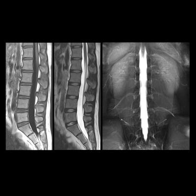
That barrier was overcome for the first time in this study by taking advantage of the fact that the 3.0T MRI scanner had a field of view long enough to image the brain and the cervical spinal cord, which relays signals to and from the hand muscles. Using this technique on subjects performing a complex finger tapping task, the authors showed that learning-related changes in blood flow in the spinal cord were independent of changes in blood flow in the brain regions involved in the task.
The results of the study indicate that the spinal cord plays an active role in the very earliest stages of motor learning. Future work will be needed to confirm that the changes seen in the spinal cord persist over time and generalize to other stages of learning and other forms of motor skills. The discovery of an independent role in learning for the spinal cord may provide new avenues for relearning motor tasks after spinal cord injury, when the connections between brain and cord are impaired.
For more information: www.plosbiology.org


 February 20, 2026


