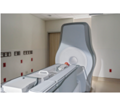
July 1, 2015 - When it comes to breast cancer screening, the fibroglandular density of your breasts affects how well a mammogram can detect cancerous tissues. That's why Pennsylvania and 20 other states have adopted laws requiring radiologists to include information about breast density in every woman's mammogram report, according to Susann Schetter, DO, co-medical director of Penn State Hershey Breast Center. Schetter's comments were published in a recent edition of The Medical Minute, a weekly health news feature produced by Penn State Milton S. Hershey Medical Center.
Breast density is described by four categories – A, B, C, and D – according to the American College of Radiology. Women who have a breast density of C (heterogeneously dense) or D (extremely dense) may want to inquire about screening methods besides mammography for early detection.
Schetter said only about 10 percent of the population has extremely fatty (A category) breasts and only about 10 percent have extremely dense (D category) breasts. Most women fall somewhere in the middle.
But breast density is not the only factor in developing breast cancer, nor is it a certain predictor of risk. For instance, younger women tend to have denser breasts, yet it is older women – who typically have less dense breast tissue – who are more at a risk for developing cancer.
And while breast density is also affected by weight, Schetter said it is always better to have a lower body-mass index (BMI) and denser breasts than to have fatty breasts and be overweight or obese because obesity increases the risk for breast cancer as well as other health problems.
"There are other metabolic activities that are going on in breast tissue that contribute to its density, so it's not just glandular tissue," she said. "We think the risk is actually highest for post-menopausal women with high breast density."
About 10 years ago, the switch to digital mammography improved identification of cancer in dense breast tissue by 30 percent, but for some women, that is still not good enough.
A mammogram image shows areas of "˜darkness,' representing fatty tissue, as well as areas of "˜whiteness' that represent the tissue which is the functional part of the breast. "The more of that white that is present on the image, the more dense the breast," Schetter said.
The problem, she said, is that cancerous tissues also show up white on a mammogram, so for women with high breast density, "it's harder for the radiologist to detect cancer because it's like looking for a snowball in a snowstorm."
Schetter said medical professionals are studying other imaging methods to supplement mammography for those with high breast density so they can do a better job of detecting cancer at the earliest stages.
Women with the highest risk of breast cancer – with more than 20 percent lifetime risk – are eligible for magnetic resonance imaging (MRI), but most women do not fall into that category. Those who have a moderately increased risk may want to consider whole-breast ultrasound, a technology that is now available at Penn State Hershey.
"It's easier to tolerate than mammography and it helps us to see inside the dense breast tissue in a different way," Schetter said. "It's also nice because an ultrasound doesn't use radiation, yet it gives us an image from the skin to chest wall of the breast tissue so we can page through and look for abnormalities."
When used in combination with mammography, the new technology is able to detect an additional two or three cancers in every 1,000 women screened. The current compromise is the possibility for false positive tests that may require a "˜second look' to determine normal breast tissue.
"So far, it has great promise," Schetter said.
For more information: www.pennstatehershey.org


 February 06, 2026


